

Diffuse cutaneous mastocytosis with generalized bullae
- PMID: 20548889
- PMCID: PMC2883405
- DOI: 10.5021/ad.2010.22.1.77
Diffuse cutaneous mastocytosis with generalized bullae
Abstract
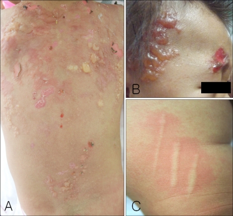
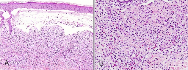
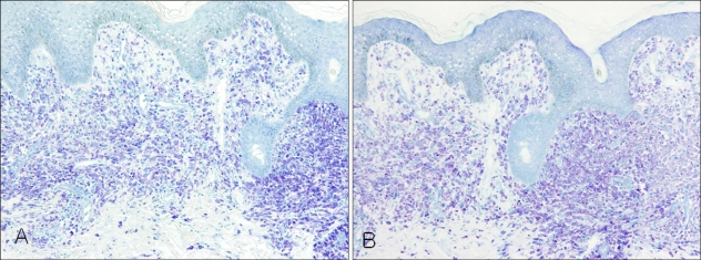
We report on a 9-month-old female infant with multiple tense bullae and erosions covering the entire body, including the face, scalp, and trunk. The histopathological examination revealed sub-epidermal bullae with a dense dermal cellular infiltrate. The infiltrate was identified as a collection of mast cells using toluidine blue and Giemsa stains. The direct immunofluorscence was negative. A diagnosis of cutaneous diffuse mastocytosis with generalized bullae was made based on these clinical and histopathological findings. In cases with diffuse cutaneous mastocytosis with generalized bullae, systemic involvement is more frequent and more severe compared to other types of cutaneous mastocytosis. Some lethal outcomes have been reported. This is the first reported case of diffuse cutaneous mastocytosis in the Korean literature.
Keywords: Diffuse cutaneous mastocytosis; Generalized bullae.
Figures
References
-
- Valent P, Horny HP, Escribano L, Longley BJ, Li CY, Schwartz LB, et al. Diagnostic criteria and classification of mastocytosis: a consensus proposal. Leuk Res. 2001;25:603–625. - PubMed
-
- Orkin M, Good RA, Clawson CC, Fisher I, Windhorst DB. Bullous mastocytosis. Arch Dermatol. 1970;101:547–564. - PubMed
-
- Longley J, Duffy TP, Kohn S. The mast cell and mast cell disease. J Am Acad Dermatol. 1995;32:545–561. - PubMed
-
- Tharp MD. Mastocytosis. In: Bolognia JL, Jorizzo JL, Rapini RP, editors. Dermatology. 2nd ed. New York: Mosby; 2008. pp. 1845–1853.
-
- Fernandez AT, Campoamor LN, Mora LE, Zambrano AZ. Diagnosis, management and classification of pediatric mastocytosis. A study of 172 cases. Actas Dermosifiliogr. 1998;89:461–476.
Publication types
LinkOut - more resources
Full Text Sources
