

Clinicopathologic manifestations of 36 korean patients with acute generalized exanthematous pustulosis: a case series and review of the literature
- PMID: 20548906
- PMCID: PMC2883418
- DOI: 10.5021/ad.2010.22.2.163
Clinicopathologic manifestations of 36 korean patients with acute generalized exanthematous pustulosis: a case series and review of the literature
Abstract
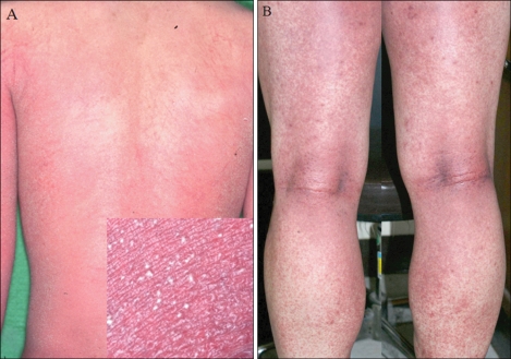
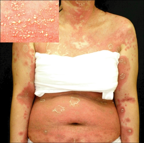
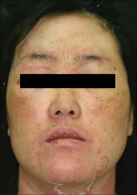
Background: Acute generalized exanthematous pustulosis (AGEP) is a rare and severe subtype of drug eruption, characterized by acute, extensive, non-follicular, sterile pustules on an erythematous background, accompanied by fever and leukocytosis.
Objective: The purpose of this study was to characterize AGEP in Korean patients in terms of clinical, laboratory, and pathologic findings.
Methods: Thirty-six patients (M:F=17:19) with AGEP were identified from an extensive review of medical records over a 15 year period. All patient cases were confirmed by biopsy and fulfilled the diagnostic criteria.
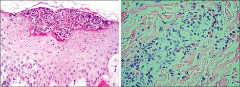
Results: The patient ages ranged from 4~80 years (37.6+/-19.4). The incubation period was 1~23 days. The duration of disease was 5~14 days. Neutrophilia (36/36), high CRP (14/36), and eosinophilia (30/36) were common laboratory findings. A history of drug administration existed in 23 of 36 patients; herbal medications, lacquers and radiocontrast media were the unique causative drugs. Spongioform subcorneal or intraepidermal pustules in the epidermis was observed in all patients. Thirty-six patients were subdivided into 2 groups: group A (n=23) was strongly associated with known agents; and group B (n=13) had no identified causative agents. There was no significant difference between the 2 groups.
Conclusion: OUR RESULTS DEMONSTRATE THE CHARACTERISTIC FEATURES OF AGEP IN KOREAN PATIENTS AS FOLLOWS: lower identification of causative agents; herbal medications, lacquers, and radiocontrast media were the main causative agents; and no significant differences existed between the 2 groups.
Keywords: Acute generalized exanthematous pustulosis; Clinicopathologic manifestation; Korean.
Figures
References
-
- Baker H, Ryan TJ. Generalized pustular psoriasis. A clinical and epidemiological study of 104 cases. Br J Dermatol. 1968;80:771–793. - PubMed
-
- Beylot C, Bioulac P, Doutre MS. Acute generalized exanthematic pustuloses (four cases) Ann Dermatol Venereol. 1980;107:37–48. - PubMed
-
- Roujeau JC, Bioulac-Sage P, Bourseau C, Guillaume JC, Bernard P, Lok C, et al. Acute generalized exanthematous pustulosis. Analysis of 63 cases. Arch Dermatol. 1991;127:1333–1338. - PubMed
-
- Chang SL, Huang YH, Yang CH, Hu S, Hong HS. Clinical manifestations and characteristics of patients with acute generalized exanthematous pustulosis in Asia. Acta Derm Venereol. 2008;88:363–365. - PubMed
-
- Rouchouse B, Bonnefoy M, Pallot B, Jacquelin L, Dimoux-Dime G, Claudy AL. Acute generalized exanthematous pustular dermatitis and viral infection. Dermatologica. 1986;173:180–184. - PubMed
LinkOut - more resources
Full Text Sources
Research Materials
Miscellaneous
