. 2010 Jun 10;6(6):e1000980.
doi: 10.1371/journal.pgen.1000980.

Cushing's syndrome and fetal features resurgence in adrenal cortex-specific Prkar1a knockout mice

Affiliations

Cushing's syndrome and fetal features resurgence in adrenal cortex-specific Prkar1a knockout mice

Isabelle Sahut-Barnola et al. PLoS Genet. .

Abstract

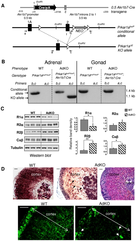
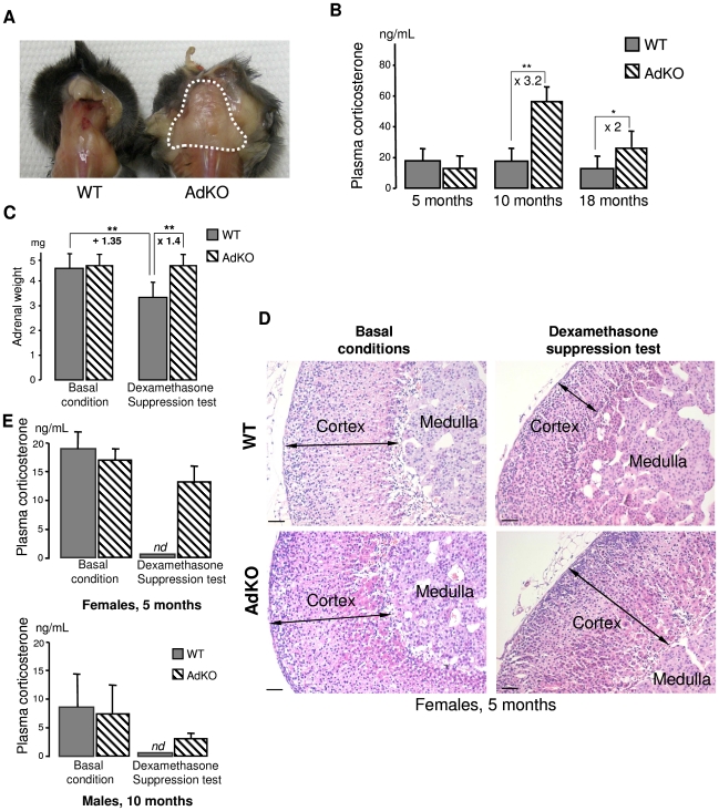
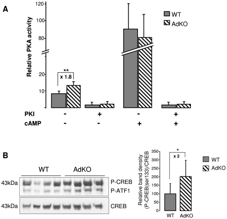
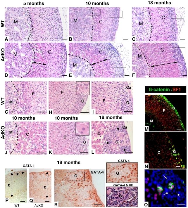
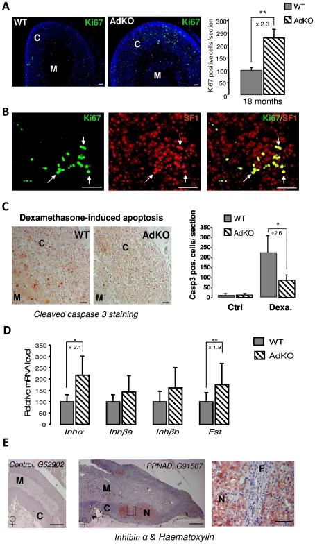
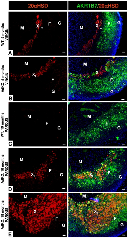
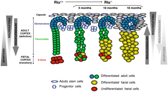
Carney complex (CNC) is an inherited neoplasia syndrome with endocrine overactivity. Its most frequent endocrine manifestation is primary pigmented nodular adrenocortical disease (PPNAD), a bilateral adrenocortical hyperplasia causing pituitary-independent Cushing's syndrome. Inactivating mutations in PRKAR1A, a gene encoding the type 1 alpha-regulatory subunit (R1alpha) of the cAMP-dependent protein kinase (PKA) have been found in 80% of CNC patients with Cushing's syndrome. To demonstrate the implication of R1alpha loss in the initiation and development of PPNAD, we generated mice lacking Prkar1a specifically in the adrenal cortex (AdKO). AdKO mice develop pituitary-independent Cushing's syndrome with increased PKA activity. This leads to autonomous steroidogenic genes expression and deregulated adreno-cortical cells differentiation, increased proliferation and resistance to apoptosis. Unexpectedly, R1alpha loss results in improper maintenance and centrifugal expansion of cortisol-producing fetal adrenocortical cells with concomitant regression of adult cortex. Our data provide the first in vivo evidence that loss of R1alpha is sufficient to induce autonomous adrenal hyper-activity and bilateral hyperplasia, both observed in human PPNAD. Furthermore, this model demonstrates that deregulated PKA activity favors the emergence of a new cell population potentially arising from the fetal adrenal, giving new insight into the mechanisms leading to PPNAD.


Conflict of interest statement

The authors have declared that no competing interests exist.

Figures

Figure 1
Figure 1. Generation of a conditional knockout for Prkar1a in mouse adrenal cortex (AdKO mice).
(A) Scheme of the 0.5 Akr1b7-Cre transgene driving the Cre expression in adrenal cortex, the Prkar1a floxed allele: Prkar1aloxP and the knockout Prkar1a allele lacking exon 2: Prkar1aΔ2. Black rectangles, exons; white triangles, loxP sites; small arrows, primers used for genotyping (see Materials and Methods for details). (B) Genomic PCR experiments using primers a, c and b, c (see A) were performed to confirm all the genotypes. Corresponding phenotypes are indicated. Intact floxed allele was amplified using primers b and c and knockout allele was amplified using primers a and c. (C) Levels of the different PKA subunits were quantified by western blotting in adrenals of 10-month-old WT vs AdKO parous female mice. * P<0.05; ** P<0.01. (D) Top panels: In situ hybridization of adrenal tissue from WT (left) and AdKO (middle and with haematoxylin/eosin staining in the right) parous female mice (10-month-old) with Prkar1a antisense riboprobe. Lower panels: R1α protein was immunodetected in adrenal sections of WT (left) and AdKO (right) 18 month-old parous females. Arrows in AdKO indicate some of the few cortical cells in which Prkar1a expression is maintained. Scale bars, 50 µm.
Figure 2
Figure 2. AdKO mice exhibited an age-dependent, dexamethasone-resistant increase in plasma corticosterone.
(A) Abnormal accumulation of adipose tissue (dotted white line) in the back of AdKO females (10-month-old). (B) Quantitative analysis of plasma corticosterone in WT female mice compared to AdKO at 5, 10 and 18 months. (C) Mean adrenal weights of 5-month-old WT and AdKO female mice (parous) in basal conditions or after 4 days dexamethasone suppression test. (D) Representative haematoxylin and eosin adrenal staining in the same conditions as (C). Scale bars, 50 µm. (E) Quantitative analysis of plasma corticosterone in basal conditions or after dexamethasone suppression test, in 5-month-old parous females and 10-month-old males WT mice compared to AdKO mice. * P<0.05; ** P<0.01.
Figure 3
Figure 3. PKA activity was increased in AdKO adrenals.
(A) In vitro quantification (arbitrary units) of the PKA catalytic activity in adrenal extracts of 10-month-old WT and AdKO females. The activity was positively or negatively regulated by cAMP, the ligand of regulatory subunits, and/or PKI, a selective inhibitor of the PKA catalytic subunits, respectively. ** P<0.01. (B) Representative CREB/P-CREB western blotting and quantification of the CREB protein phosphorylation in 10 months WT and AdKO adrenals. * P<0.05.
Figure 4
Figure 4. Morphological defects and progressive hyperplasia in AdKO adrenals.
(A–L) Adrenals were collected from WT (A–C and G–I) and AdKO (D–F and J–L) parous females aged of 5, 10 and 18 months and stained with haematoxylin and eosin. AdKO adrenal cortex presented progressive centrifugal expansion of large eosinophilic cells, indicated by a double arrow in (D–F). In the top panels, squares delineate the higher magnifications shown in (G–I) for WT and (J–L) for AdKO adrenals. Insets, higher magnification illustrating the increased cell area of expanding eosinophilic cells compared to normal spongiocytes. (M–O) Adrenal sections from 18-month-old mice double-stained for β-catenin, a zona glomerulosa marker (in green) and for Sf1, a steroidogenic marker (in red). Nuclei were stained by Hoechst in blue. (M) WT adrenal cortex. (N) AdKO adrenal cortex. (O) Higher magnification of (N), where hyperplastic small spindle-shaped, basophilic cells lacking both staining are enlightened by arrows. (P–R) Immunodetection of the pre-tumoural marker Gata-4 in adrenal sections of parous 18-month-old females. (P) WT. (Q–R) AdKO. (R) The square delineates the higher magnifications shown in the right. Top panel: Gata-4 staining. Lower panel: same section with haematoxylin and eosin counter-staining. M, Medulla; C, Cortex; F, zona fasciculata; G, zona glomerulosa; Ca, Capsule; Scale bars, 50 µm.
Figure 5
Figure 5. Proliferation and resistance to apoptosis in AdKO adrenals, inhibin-activin system in AdKO adrenals, and PPNAD.
(A) Adrenals from 18-month-old WT and AdKO parous females were immunodetected with Ki67 proliferation marker. The corresponding statistical analysis of the number of Ki67 expressing cells in the adrenal cortex of both genotypes is shown on the right panel. ** P<0.01. (B) Co-immunodetection of Ki67 (green) and Sf1 (steroidogenic marker, red) in 18 months AdKO adrenal sections. Co-localisation of Ki67 and Sf1 stainings is shown in the right panel. (C) Representative stainings for cleaved-caspase 3 apoptosis marker in WT and AdKO adrenal sections of 5-month-old mice treated for 4 days with dexamethasone. The right panel shows the quantification of apoptotic cells (cleaved-caspase 3 positive) in adrenal cortex of untreated (Ctrl) or treated (Dexa) mice of both genotypes. * P<0.05. (D) Quantitative representation of mRNA expression (RT-QPCR) of genes encoding inhibin-activin system in 10-month-old parous female adrenals of both genotypes. Inhibin subunit (Inhα); Activin A and B subunits (Inhβa and Inhβb); Follistatin (Fst). * P<0.05; ** P<0.01. (E) Immunostaining for INHIBIN-α on human adrenal sections from control G52902 patient (female) and PPNAD G91567 patient (female) carrying germline PRKAR1A-inactivating mutation c.709-7del6(TTTTTA) . Similar stainings were observed on two other control patients (not shown) and four other PPNAD patients (Figure S6). Square delineates the higher magnifications located on the right. N, nodule; M, medulla, F, zona fasciculata. Scale bars, 15 mm in entire human adrenal and 50 µm in magnification.
Figure 6
Figure 6. Existence of a persistent, mislocated X-like-zone in AdKO adrenals.
The X-zone 20α-HSD marker (in red) and the zona fasciculata Akr1b7 marker (green, right column) were co-immunodetected. The two colours are merged in the right column with the Hoechst nuclei marker (blue). (A) Adrenal section of a 5-month-old nulliparous WT female. The unlabelled zone separating the X-zone (red) and the zona fasciculata (green) is indicated by a star (*). (B) Adrenal section of a 5-month-old nulliparous AdKO female. (C) Adrenal section of a 10-month-old parous WT female. (D) Adrenal section of a 10-month-old parous AdKO female. (E) Adrenal section of a 18-month-old parous AdKO female. M, Medulla; F, zona fasciculata; G, zona glomerulosa; X, X-zone; XL, X-like-zone; Scale bars, 50 µm.
Figure 7
Figure 7. AdKO adrenals expressed Cyp17 and produced cortisol.
All the experiments were realised in 10-month-old parous females. (A) Quantitative representation (RT-QPCR) of Cyp17 mRNA levels, a foetal adrenal marker in mouse, in WT and AdKO adrenals. (B) Representative immunodetection of Cyp17 showing abnormal expression in the innercortex in AdKO compared to WT adrenals. (C) Quantitative analysis of plasma cortisol showing significant level of cortisol in AdKO compared to normal absence in WT mice. * p<0.05.
Figure 8
Figure 8. Model of dynamic changes and cell renewal in the AdKO adult cortex.
Left, model of cell renewal and homeostatic growth maintenance of WT adrenal cortex (according to [17]). Hypothetical opposing gradients of proliferation and apoptosis leading to the set up of centripetal differentiation are schematized. Right, consequences of the defect of apoptosis gradient on the cellular dynamics in AdKO cortex (5-, 10-, and 18-month-old mice). Loss of R1α led to decreased apoptosis of foetal cells which were maintained, expanded across the cortex and differentiated (acquired steroidogenic competences) while definitive cortex progressively regressed.

Similar articles

Cited by

References

    1. Carney JA, Gordon H, Carpenter PC, Shenoy BV, Go VL. The complex of myxomas, spotty pigmentation, and endocrine overactivity. Medicine (Baltimore) 1985;64:270–283. - PubMed
    1. Kirschner LS, Carney JA, Pack SD, Taymans SE, Giatzakis C, et al. Mutations of the gene encoding the protein kinase A type I-alpha regulatory subunit in patients with the Carney complex. Nat Genet. 2000;26:89–92. - PubMed
    1. Casey M, Vaughan CJ, He J, Hatcher CJ, Winter JM, et al. Mutations in the protein kinase A R1alpha regulatory subunit cause familial cardiac myxomas and Carney complex. J Clin Invest. 2000;106:R31–38. - PMC - PubMed
    1. Groussin L, Jullian E, Perlemoine K, Louvel A, Leheup B, et al. Mutations of the PRKAR1A gene in Cushing's syndrome due to sporadic primary pigmented nodular adrenocortical disease. J Clin Endocrinol Metab. 2002;87:4324–4329. - PubMed
    1. Amieux PS, Howe DG, Knickerbocker H, Lee DC, Su T, et al. Increased basal cAMP-dependent protein kinase activity inhibits the formation of mesoderm-derived structures in the developing mouse embryo. J Biol Chem. 2002;277:27294–27304. - PubMed

Publication types

MeSH terms

Substances