. 2010 Jun 10;6(6):e1000948.
doi: 10.1371/journal.ppat.1000948.

A crucial role for infected-cell/antibody immune complexes in the enhancement of endogenous antiviral immunity by short passive immunotherapy

Affiliations

A crucial role for infected-cell/antibody immune complexes in the enhancement of endogenous antiviral immunity by short passive immunotherapy

Henri-Alexandre Michaud et al. PLoS Pathog. .

Abstract

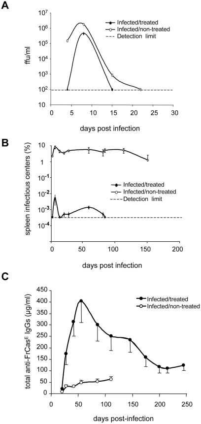
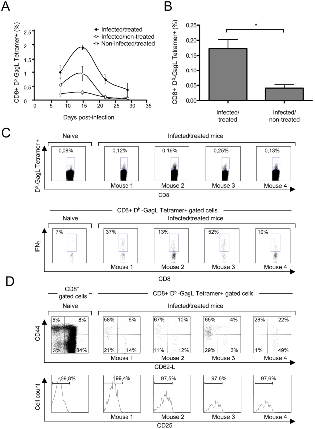
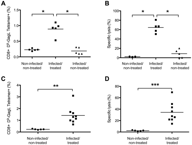
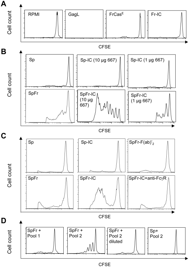
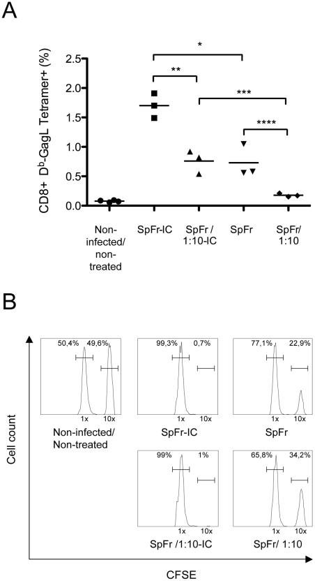
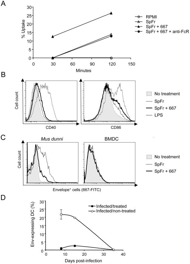
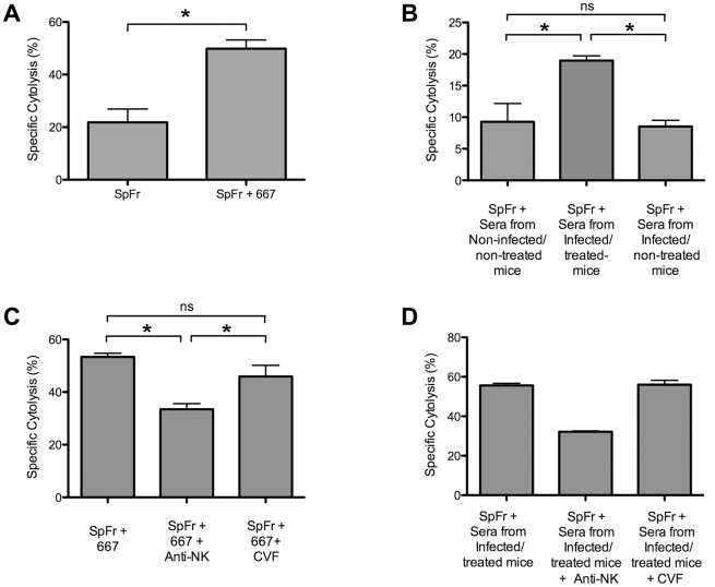
Antiviral monoclonal antibodies (mAbs) represent promising therapeutics. However, most mAbs-based immunotherapies conducted so far have only considered the blunting of viral propagation and not other possible therapeutic effects independent of virus neutralization, namely the modulation of the endogenous immune response. As induction of long-term antiviral immunity still remains a paramount challenge for treating chronic infections, we have asked here whether neutralizing mAbs can, in addition to blunting viral propagation, exert immunomodulatory effects with protective outcomes. Supporting this idea, we report here that mice infected with the FrCas(E) murine retrovirus on day 8 after birth die of leukemia within 4-5 months and mount a non-protective immune response, whereas those rapidly subjected to short immunotherapy with a neutralizing mAb survive healthy and mount a long-lasting protective antiviral immunity with strong humoral and cellular immune responses. Interestingly, the administered mAb mediates lysis of infected cells through an antibody-dependent cell cytotoxicity (ADCC) mechanism. In addition, it forms immune complexes (ICs) with infected cells that enhance antiviral CTL responses through Fc gammaR-mediated binding to dendritic cells (DCs). Importantly, the endogenous antiviral antibodies generated in mAb-treated mice also display the same properties, allowing containment of viral propagation and enhancement of memory cellular responses after disappearance of the administered mAb. Thus, our data demonstrate that neutralizing antiviral mAbs can act as immunomodulatory agents capable of stimulating a protective immunity lasting long after the end of the treatment. They also show an important role of infected-cells/antibody complexes in the induction and the maintenance of protective immunity through enhancement of both primary and memory antiviral T-cell responses. They also indicate that targeting infected cells, and not just viruses, by antibodies can be crucial for elicitation of efficient, long-lasting antiviral T-cell responses. This must be considered when designing antiviral mAb-based immunotherapies.


Conflict of interest statement

The authors have declared that no competing interests exist.

Figures

Figure 1
Figure 1. Differences in viremia, spleen infectious centers and anti-FrCasE IgG responses between infected/treated and infected/non-treated mice.
Serum viremia, presented as focus-forming units (ffu)/ml (A) and spleen infectious centers (B) were assayed as described in the Methods section. Two groups of 8 day-old mice were infected with FrCasE. One group was treated with 667 (infected/treated) 1 hour post-infection and on days 2 and 5 post-infection and the other was not treated (infected/non-treated). A group of mice that was not infected but that was treated with 667 (non-infected/treated), was taken as a negative control (not shown). The data presented are representative of 2 independent experiments. Each time point is the average of values obtained from 2 animals per time point except in the case of day 8 (5 animals). Non-infected/treated mice were negative for both serum viremia and SICs and are not presented. Error bars indicate SEM. Total anti-FrCasE IgGs (C) were assayed by ELISA. Mice were infected and treated as in A and B. One of 2 independent experiments with similar outcomes is presented. The data are the average of values obtained from at least 10 animals per time point. Non-infected/treated mice were negative for anti-FrCasE IgGs and are therefore not presented. Error bars indicate SEM.
Figure 2
Figure 2. Primary CD8+ T-cell responses against FrCasE-infected cells.
Mice were infected and treated, or not, as in Figure 1 . (A) Kinetics of the primary anti-FrCasE CTL response. Spleen cells from 2 mice of each group per time point (days 8, 22 and 28 post-infection) and from 5 mice per group at day 15 post-infection were stained with an anti-CD8 mAb and the Db-GagL tetramer and analyzed by flow cytometry. (B–D) Phenotypic characterization of primary CD8+ T-cell responses in infected/treated animals. Mice were infected and treated as in Figure 1 . CD3+ cells were isolated by negative selection from the spleens of 4 infected/treated- and 2 infected/non-treated mice on day 56 post-infection. Half of the CD3+ cells were then stained with the Db-GagL tetramer and anti-CD8, anti-CD44, anti-CD62L and anti-CD25 mAb and analyzed by flow cytometry. In parallel, the other half of the CD3+ cells were incubated with PMA-ionomycin plus brefeldin A and IFN-γ production by tetramer+CD8+ T cells was assessed by intracellular staining. The statistical significance of data between infected/treated- and infected/non-treated mice was established using the Student's t test (*P = 0,045). (B) Assay of GagL-specific CD8+ T cells in infected/treated and infected/non-treated mice on day 56 post-infection. (C–D) Phenotypic characterization of GagL-specific CD8+ T cells on day 56 post-infection. Data from 1 age-matched non-infected/non-treated (naive) mouse and 4 infected/treated mice are presented.
Figure 3
Figure 3. Long-term CTL memory responses in infected/treated animals versus control mice.
Mice were challenged with FrCasE-infected splenocytes on days 56 or 116 post-infection and 9 days later CD8+ T-cell responses were assessed by measuring both the percentage of GagL-specific CD8+ T cells and specific in vivo CTL activity as described in the Methods section. (A–B) Assay of GagL-specific memory CTL responses in infected/treated and infected/non-treated mice challenged on days 56 or 116 post-infection. Infected/treated and infected/non-treated mice were challenged on day 56 (3 mice per group) or on day 116 (2 mice per group). Non-challenged age-matched non-infected/non-treated mice (3 on day 56 and 2 on day 116) were used as controls. The statistical significance of data between the infected/treated group and the other two groups was established using a non-parametric one-way ANOVA test with Dunn's multiple comparison post-test (*P<0,05). (C–D) Assay of GagL-specific memory CTL responses in infected/treated mice challenged at month 8 post-infection. Eight month-old infected/treated mice were challenged with FrCasE-infected splenocytes. Non-challenged aged-matched non-infected/non-treated mice were used as controls. Statistical significance of data between infected/treated- and non-infected/non-treated mice was established using the Student's t test (**P = 0,0034; *** P = 0,0015). Bars indicate mean values.
Figure 4
Figure 4. Stimulation of proliferation of GagL-TCR-TG CD8+ T cells by 667 and sera from infected/treated mice.
BMDCs were prepared and exposed to various sources of antigen. The day after, they were co-cultured with CSFE-labeled CD8+ T cells prepared from lymph nodes from GagL-TCR-TG mice transgenic for a TCR recognizing the H2Db-restricted GagL peptide. Proliferation was assessed 5 days later by flow cytometry. (A) Control experiments. BMDCs were cultivated with the GagL peptide (used as positive control), FrCasE and FrCasE ICs made with 667 (Fr-IC). Culture medium (RPMI) served as a negative control. (B) Stimulation of CD8+ T cells priming by FrCasE-infected splenocytes in the presence and in the absence of 667. Non-infected (Sp) or infected splenocytes (SpFr) were incubated in the presence of 2 concentrations of 667 (Sp-IC and SpFr-IC, respectively). A typical experiment out of 3 independent ones with similar outcomes is presented. The statistical significance between the SpFr and SpFr-IC groups in these 3 experiments was established using the Student's t test (*P = 0,0003) (see Figure S4). (C) Stimulation of CD8+ T cells priming by FrCasE-infected splenocytes complexed with either the 667 F(ab')2 fragment or the whole 667 mAb in the presence and in the absence of the FcγR-blocking antibody. FrCasE-infected splenocytes (SpFr) were complexed with either 15 µg of the 667 F(ab')2 (SpFr- F(ab')2) or with 10 µg of the 667 mAb (SpFr-IC). SpFr-IC were co-cultured with BMDCs previously incubated (SpFr-IC + anti-FcγR), or not, with the FcγR-blocking antibody. Non-infected splenocytes incubated in the absence (Sp) or in the presence of 10 µg of 667 (Sp-IC) were used as control. (D) Stimulation of CD8+ T cells priming by FrCasE-infected splenocytes in the presence of sera from infected/treated mice or infected/non-treated mice. Pool 1 corresponds to sera from 4 infected/non-treated animals sacrificed on day 115 post-infection whereas Pool 2 corresponds to the sera of 4 infected/treated mice sacrificed on day 243. “Pool 2-diluted” corresponds to Pool 2 with anti-FrCasE antibody concentration adjusted to that of Pool 1. Similar results were obtained in 2 independent experiments with individual serum samples from mice of each group.
Figure 5
Figure 5. In vivo enhancement of CD8+ T-cell responses against FrCasE by ICs.
SpFr or SpFr-IC were administered i.v. to non-infected/non-treated mice. Four groups of mice (n = 3) where used: two of them were injected with either 2×10⁶ or 2×10⁵ FrCasE-infected splenocytes (SpFr and SpFr 1:10, respectively) in the absence of 667, and the other two were injected with the same amount of FrCasE-infected splenocytes complexed to 150 µg of 667 (SpFr-IC and SpFr 1:10-IC respectively). Nine days later, they were tested for the expansion of GagL-specific CD8+ T cells and for CTL activity in vivo as described in Figure 3 . Non-infected/non-treated mice with no further treatment were used as controls (A). Proliferation of GagL-specific CD8+ cells. The statistical significance between groups was established using the Student's t test (*P = 0,0106; **P = 0,0049; ***P = 0,0071: ****P = 0,0352). (B) CTL activity against GagL-loaded splenocytes. A representative mouse of each group is presented.
Figure 6
Figure 6. Influence of FrCasE-infected cell ICs on BMDCs.
(A) Uptake of SpFr and SpFr-IC by DCs. PKH26-labelled SpFr or -SpFr-IC were given to BMDCs and uptake was quantified by flow cytometry in the presence, or in the absence, of the 2.4G2 FcγR-blocking mAb. One experiment out of 3 with similar outcomes is presented. (B) BMDCs activation. BMDCs were incubated for 24 hours in the presence of SpFr, SpFr-IC or LPS before flow cytometry assay of CD40 and CD86. One representative experiment out of 3 independent with similar outcomes is presented. (C) Assessment of productive in vitro BMDC infection. BMDC were co-cultured with SpFr or SpFr-IC for 48 hours before flow cytometry assay of MHC-II+Env+ cells. Infection-sensitive Mus dunni fibroblasts were used as positive controls in parallel experiments. (D). Assay of Env-expressing DCs in vivo. MHC-II+Env+ splenocytes from infected/treated- and infected/non-treated mice were assayed by flow cytometry at days 8, 15 and 35 post-infection.
Figure 7
Figure 7. Cytolytic activity against FrCasE-infected cells of 667 and of anti-FrCasE antibodies endogenously produced in infected/treated mice.
In vivo cytolysis activities were assayed after administration of CFSE-labeled FrCasE-infected splenocytes (SpFr) followed by administration of antibodies from various sources and cytometry analysis after 5 or 24 hours. (A) Cytolytic activity of 667. SpFr were administered i.v. and, then, 667 or PBS i.p. Values are the average ± SEM of experiments conducted with 4 mice per condition. The statistical significance was established using the Student's t test (*P = 0,0007). (B) Cytolytic activity of sera from infected/treated- and infected/non-treated mice. After administration of SpFr, three groups of 4 mice each were treated with equivalent volumes (175 µl) of pools of sera from 2 non-infected/non-treated-, 2 infected/non-treated- and 2 infected/treated mice. Values are the average ± SEM of experiments conducted with 4 mice per condition. Statistical significance of sera from infected/treated group versus the other two groups was determined by a non-parametric one-way ANOVA test with Dunn's multiple comparison post-test. *P<0,05. (C) Cytolytic activity of 667 in mice treated with either anti-NK antibodies or cobra venom factor. SpFr mixed with 667, or PBS for control, were administered i.v. to mice treated or not with either anti-NK antibodies or the cobra venom factor as described in the Methods section. The values are the average ± SEM of experiments conducted with 7 mice per condition. The statistical significance of the data obtained with sera from the infected/treated group versus those obtained in the other two groups was established using a non-parametric one-way ANOVA test with Dunn's multiple comparison post-test. (*P<0,05). (D) Cytolytic activity of sera from infected/treated in mice treated with anti-NK antibodies or cobra venom factor. SpFr mixed to 175 µl of pools of sera from 4 infected/treated were administered i.v. to mice treated or not with either anti-NK antibodies or the cobra venom factor, as described in the Methods section. The values are the average ± SEM of experiments conducted with 2 mice per condition. Statistical significance of sera from infected/treated group versus the other two groups was not assessed due to the reduced number of mice per condition.

Similar articles

Cited by

References

    1. Reichert JM. Trends in the development and approval of monoclonal antibodies for viral infections. BioDrugs. 2007;21:1–7. - PMC - PubMed
    1. Marasco WA, Sui J. The growth and potential of human antiviral monoclonal antibody therapeutics. Nat Biotechnol. 2007;25:1421–1434. - PMC - PubMed
    1. Takada A, Ebihara H, Jones S, Feldmann H, Kawaoka Y. Protective efficacy of neutralizing antibodies against Ebola virus infection. Vaccine. 2007;25:993–999. - PubMed
    1. Oliphant T, Nybakken GE, Engle M, Xu Q, Nelson CA, et al. Antibody recognition and neutralization determinants on domains I and II of West Nile Virus envelope protein. J Virol. 2006;80:12149–12159. - PMC - PubMed
    1. Hangartner L, Zinkernagel RM, Hengartner H. Antiviral antibody responses: the two extremes of a wide spectrum. Nat Rev Immunol. 2006;6:231–243. - PubMed

Publication types

MeSH terms