. 2010 Jun 10;6(6):e1000982.
doi: 10.1371/journal.pgen.1000982.

Arabidopsis thaliana chromosome 4 replicates in two phases that correlate with chromatin state

Affiliations

Arabidopsis thaliana chromosome 4 replicates in two phases that correlate with chromatin state

Tae-Jin Lee et al. PLoS Genet. .

Abstract

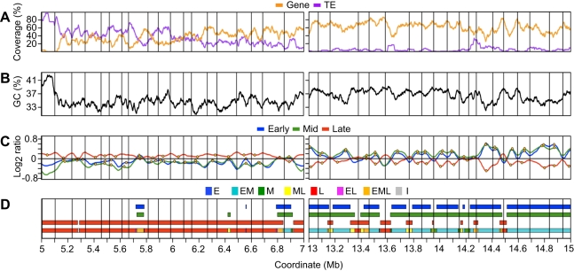
DNA replication programs have been studied extensively in yeast and animal systems, where they have been shown to correlate with gene expression and certain epigenetic modifications. Despite the conservation of core DNA replication proteins, little is known about replication programs in plants. We used flow cytometry and tiling microarrays to profile DNA replication of Arabidopsis thaliana chromosome 4 (chr4) during early, mid, and late S phase. Replication profiles for early and mid S phase were similar and encompassed the majority of the euchromatin. Late S phase exhibited a distinctly different profile that includes the remaining euchromatin and essentially all of the heterochromatin. Termination zones were consistent between experiments, allowing us to define 163 putative replicons on chr4 that clustered into larger domains of predominately early or late replication. Early-replicating sequences, especially the initiation zones of early replicons, displayed a pattern of epigenetic modifications specifying an open chromatin conformation. Late replicons, and the termination zones of early replicons, showed an opposite pattern. Histone H3 acetylated on lysine 56 (H3K56ac) was enriched in early replicons, as well as the initiation zones of both early and late replicons. H3K56ac was also associated with expressed genes, but this effect was local whereas replication time correlated with H3K56ac over broad regions. The similarity of the replication profiles for early and mid S phase cells indicates that replication origin activation in euchromatin is stochastic. Replicon organization in Arabidopsis is strongly influenced by epigenetic modifications to histones and DNA. The domain organization of Arabidopsis is more similar to that in Drosophila than that in mammals, which may reflect genome size and complexity. The distinct patterns of association of H3K56ac with gene expression and early replication provide evidence that H3K56ac may be associated with initiation zones and replication origins.


Conflict of interest statement

The authors have declared that no competing interests exist.

Figures

Figure 1
Figure 1. Flow cytometric analysis of Arabidopsis cell suspension culture.
(A) Analytical FACS profile showing BrdU incorporation into Arabidopsis nuclei as a function of DNA content. BrdU incorporation and DNA content of nuclei were visualized using anti-BrdU Alexa 488 conjugate and propidium iodide, respectively. Five boxes are shown, representing nuclei in G1, early S, mid S, late S, and G2/M, respectively. (B) Histogram plots for total and BrdU-positive nuclei from the BrdU labeled cells shown in (A). (C) Flow diagram of FACS-based microarray experiments for profiling replication in early, mid and late S. Cells were pulse-labeled with BrdU, and nuclei isolated. Populations of nuclei in early S/G1, mid S, and late S/G2 were sorted based on DNA content. Genomic DNA was prepared from the sorted nuclei in each fraction and sheared to an average size of 500 bp before heat denaturation and immunoprecipitation with antibodies against BrdU. DNA containing BrdU was amplified, labeled with Cy dyes and hybridized to a tiling array for Arabidopsis chr 4.
Figure 2
Figure 2. Arabidopsis chromosome 4 with replication profiles for early, mid, and late S phase suspension culture cells.
(A) Gene and TE coverage were determined using TAIR8 annotation and are expressed as percentage of bases in 1 kb non-overlapping segments occurring in genes or TEs respectively. Overlapping genes or TEs were merged to prevent coverage values exceeding 100%. Data were loess-smoothed using a 150 kb window. (B) GC percentage calculated in 1 kb non-overlapping windows and loess-smoothed using a 150 kb window. (C) Schematic representation of chromosome 4 omitting the telomeres and nucleolar organizing region. The gene-rich euchromatic distal short and distal long arms are shaded light gray while the heterochromatic knob and pericentromere are rich in TEs and are shaded black. The proximal portions of both the short and long arms have intermediate characteristics and are shaded dark gray. (D) Replication profiles for early, mid, and late S phase cells. Replication is expressed as log2 ratio of BrdU-labeled sequences in early, mid or late S phase cells with respect to total DNA from the same cells. Data have been normalized and scaled within experiments, but no normalization was performed between experiments.
Figure 3
Figure 3. Analysis of replication profiles for segments of coordinate replication time and identification of initiation and termination zones.
Representative late- (coordinates 5–7 Mb) and early-replicating (coordinates 13–15 Mb) regions of chromosome 4 are shown. (A) Gene coverage and TE coverage as in Figure 2 but the window for loess-smoothing is 50 kb. Vertical gridlines across all panels are described in (C). (B) GC percentage loess-smoothed using a 50 kb window. (C) Replication profiles as in Figure 2 for early, mid, and late S phase cells. Putative initiation zones are shown as orange circles while putative termination zones are shown as vertical gridlines. (D) Segmentation of replication profiles into segments of coordinate timing. Segments of early, mid, or late replication timing were identified within each experiment as regions where the loess-smoothed profile showed enrichment for BrdU-labeling (log2 ratio >0). A final step was performed to reconcile timing between experiments and to account for the overlap of segments between experiments, as shown in the composite model (lowest line). Segments were classified as E (only early), EM (early and mid), M (only mid), ML (mid and late), L (only late), EL (early and late), EML (early, mid and late) or I (indeterminate, no enrichment in any experiment).
Figure 4
Figure 4. Analysis of chromosome 4 replication timing segments and replicon structure.
(A) Distribution of replication timing segments for chr4. The majority of chr4 replicates as either EM (37%) or L segments (44%). The heterochromatic knob and pericentromere replicate almost exclusively as L segments while EM segments dominate the gene-rich distal long arm. The distal short, proximal short and proximal long arms display complex replication timing patterns intermediate between the heterochromatic and euchromatic regions. (B) Distribution and timing of replication initiation and termination zones. Replication timing was assigned to all zones from the timing of overlapping probes. The occurrence of initiation zones in EM and L segments is proportional to the coverage of EM and L segments. In contrast, termination zones are overrepresented in L segments. (C) Schematic representation of replicons, replication timing, and replication domains for chr4. In the top panel, each vertical bar represents a replicon with the width of the bar proportional to the length of the replicon. Subdivisions within the bar indicate the percentage of probes within the replicon that replicate in a given time window. The middle panel illustrates the clustering of replicons with similar timing into replication domains. For reference, the lower panel shows the chromosomal zones defined in Figure 4C. (D) Analysis of replicon size as a function of replication timing. 70 EM, 2 M, 3 ML, 80 L and 8 EML replicons were identified for chr4. Lengths of identified replicons are shown as a boxplot with whiskers extending to 1.5 times the interquartile range. Outliers are shown as open cirlces. The differences in size between replicon classes were not statistically significant as determined by a t-test.
Figure 5
Figure 5. Distribution of genetic and epigenetic features within replicons.
The proportion of AT-rich and gene-rich probes and probes positive for H3K56ac, H3K4me1/2, H3K9me3 and 5mC were calculated for each EM or L replicon in 10% intervals and binned according to position in the replicon as described in the text. Bin 1 represents the innermost 20% of the replicon while bin 5 covers the outermost 20% proximal to the termination zones. The barplots show the mean proportion in each bin for EM and L replicons. Error bars indicate the 95% confidence intervals as determined from the binomial distribution. The dashed horizontal lines indicate the mean proportion across the replicons regardless of position.
Figure 6
Figure 6. Heat maps of epigenetic modifications and gene expression.
Representative late- and early-replicating regions as in Figure 4 are shown. Loess-fitted probe enrichment values for H3K56ac, H3K4me1/2, 5mC and H3K9me2 are presented as heat maps. Gene expression is presented similarly and corresponds to loess-fitted gcRMA expression values with grey regions indicating genes not present on the ATH1 array while green regions indicate intergenic probes. The early S phase replication profile is shown to indicate replication time. H3K56ac is elevated in large regions near initiation zones while H3K4me1/2 and 5mC are enriched in large regions near termination zones, but there are no consistent correlations between gene expression, replication timing, and epigenetic modifications on this scale.

Similar articles

Cited by

References

    1. Berezney R, Dubey DD, Huberman JA. Heterogeneity of eukaryotic replicons, replicon clusters, and replication foci. Chromosoma. 2000;108:471–484. - PubMed
    1. Gilbert DM. In search of the holy replicator. Nat Rev Mol Cell Biol. 2004;5:848–855. - PMC - PubMed
    1. Aladjem MI. Replication in context: dynamic regulation of DNA replication patterns in metazoans. Nat Rev Genet. 2007;8:588–600. - PubMed
    1. Hamlin JL, Mesner LD, Lar O, Torres R, Chodaparambil SV, et al. A revisionist replicon model for higher eukaryotic genomes. J Cell Biochem. 2008;105:321–329. - PMC - PubMed
    1. Gondor A, Ohlsson R. Replication timing and epigenetic reprogramming of gene expression: a two-way relationship? Nat Rev Genet. 2009;10:269–276. - PubMed

Publication types