. 2009 Nov 21;2(4):486-494.
doi: 10.1007/s12195-009-0094-2.

INSIGHTS INTO THE ROLES OF NON-MUSCLE MYOSIN IIA IN HUMAN KERATINOCYTE MIGRATION

Affiliations

INSIGHTS INTO THE ROLES OF NON-MUSCLE MYOSIN IIA IN HUMAN KERATINOCYTE MIGRATION

Saheli Sarkar et al. Cell Mol Bioeng. .

Abstract

Epidermal cell migration is a key factor in wound healing responses, regulated by the F-actin-myosin II systems. Previous reports have established the importance of non-muscle myosin II (NMII) in regulating cell migration. However, the role of NMII in primary human keratinocytes has not been investigated. In this study we used a microfabrication-based two-dimensional migration assay to examine the role of NMII in keratinocyte migration. We developed confluent cell islands of various sizes (0.025 - 0.25 mm(2)) and quantified migration as Fold Increase in island area over time. We report here that NMII was expressed and activated in migrating keratinocytes. Inhibition of NMIIA motor activity with blebbistatin increased migration significantly in all cell island sizes in six hours compared to control. Inhibition of Rho-kinase by Y-27632 did not alter migration while inhibition of myosin light chain kinase by ML-7 suppressed migration significantly in six hours. Both blebbistatin and Y-27632 induced formation of large membrane ruffles and elongated tails. In contrast, ML-7 blocked cell spreading, resulting in a rounded morphology. Taken together, these data suggest that NMIIA decreases migration in keratinocytes, but the mechanism may be differentially regulated by upstream kinases.


Figures

Figure 1
Figure 1
Keratinocyte island migration on plastic on fibronectin (Fn). (A) Keratinocyte cell island array at t = 0 hrs. The islands shown here are square islands of size 0.025 mm2. (B) Individual square islands of sizes 0.0625 mm2, 0.125 mm2 and 0.25 mm2 at t = 0 hrs. (C) A typical 0.25 mm2 cell island after 6 hrs of migration on untreated (control) and fibronectin - coated (Fn) surface. Scale bar: 100µm.
Figure 2
Figure 2
Quantification of island migration on untreated surfaces and fibronectin (Fn). Migration is defined as Fold Increase in island area (at t = 6 hrs) normalized to the initial island area (at t = 0 hrs). Cells on Fn migrated significantly more compared to control. Data is presented as mean Fold Increase in island area ± SEM for n = 9 islands from 3 experiments.
Figure 3
Figure 3
Expression and activation of Non-Muscle Myosin IIA in non-migratory and migratory keratinocytes. (A) Expression of NMIIA and F-actin in non-migrating keratinocytes. Cell islands developed on Fn were fixed with PDMS membranes in situ (at t = 0 hrs). The cells were labeled with the following: NMIIA (green)/phalloidin (red) or pRLC (green)/phalloidin (red). Top: NMIIA was distributed diffusely in the cytosol. Bottom: pRLC expression was not found in these cells. F-actin distribution is also diffuse in the cells. (B) Expression of NMIIA and F-actin in migrating keratinocytes. After 6 hrs of migration on Fn, cells were fixed and labeled with NMIIA (green)/phalloidin (red) or pRLC (green)/phalloidin (red). Top: NMIIA was recruited to the edges in migrating cells (block arrow). Bottom: pRLC expression (indicated by block arrow) was observed specifically at the cellular edges not in contact with other cells (cell-cell contact area indicated by line arrow). F-actin is localized at the cellular edges. Cell nuclei were labeled with DAPI (blue). Scale bar: 20µm.
Figure 4
Figure 4
Effect of inhibitors on island migration. (A) Confluent cell islands were treated with blebbistatin (30 µM), ML-7 (10 µM) or Y-27632 (5 µM) as described in Materials and Methods. Migration of a typical 0.25 mm2 cell island on Fn at 6 hrs is shown here for each condition. Fn (control) indicates cells grown on fibronectin without any inhibitors. Scale bar: 100µm. (B) Quantification of island migration (at t = 6 hrs) on Fn induced by inhibitors. Migration is shown as normalized Fold Increase in island area after 6 hrs. Blebbistatin induced significant increase in keratinocytes migration (* Indicates p<0.05 compared to control).
Figure 5
Figure 5
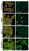
Impact of inhibitors on NMIIA localization and phosphorylation (t = 6 hrs). The left panels show visualization of F-actin (red), NMIIA (green) and DAPI (blue). The right panels show pRLC (green) and DAPI. (A) Control cells grown on Fn without any inhibitors. (B) Blebbistatin-treated cells showed large F-actin rich ruffles (block arrow) followed by polarized NMIIA bands (*) and actomyosin-rich tails (line arrow). pRLC expression was observed at the cell edges (arrowhead). (C) ML-7- treated cells showed narrow F-actin bands at the cell edges and diffuse NMIIA staining. pRLC expression was very low. (D) Y-27632 treated cells showed smaller F-actin- rich membrane ruffles (block arrow) with NMIIA distributed through the cell body (*) and some polarization toward the posterior edge of the cell. pRLC expression was strongly localized at the cell centre and trailing edges of the cells (arrowhead). Scale bar: 20µm.

Similar articles

Cited by

References

    1. Amano M, Ito M, Kimura K, Fukata Y, Chihara K, Nakano T, Matsuura Y, Kaibuchi K. Phosphorylation and activation of myosin by Rho-associated kinase (Rho-kinase) J Biol Chem. 1996;271(34):20246–20249. - PubMed
    1. Babbin BA, Koch S, Bachar M, Conti MA, Parkos CA, Adelstein RS, Nusrat A, Ivanov AI. Non-muscle myosin IIA differentially regulates intestinal epithelial cell restitution and matrix invasion. Am J Pathol. 2009;174(2):436–448. - PMC - PubMed
    1. Betapudi V, Licate LS, Egelhoff TT. Distinct roles of nonmuscle myosin II isoforms in the regulation of MDA-MB-231 breast cancer cell spreading and migration. Cancer Res. 2006;66(9):4725–4733. - PubMed
    1. Conti MA, Adelstein RS. Nonmuscle myosin II moves in new directions. J Cell Sci. 2008;121(Pt 1):11–18. - PubMed
    1. Duxbury MS, Ashley SW, Whang EE. Inhibition of pancreatic adenocarcinoma cellular invasiveness by blebbistatin: a novel myosin II inhibitor. Biochem Biophys Res Commun. 2004;313(4):992–997. - PubMed

LinkOut - more resources