

The emerging clinical role of cardiovascular magnetic resonance imaging
- PMID: 20548977
- PMCID: PMC2903987
- DOI: 10.1016/s0828-282x(10)70396-2
The emerging clinical role of cardiovascular magnetic resonance imaging
Abstract
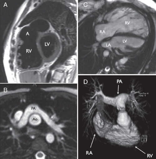
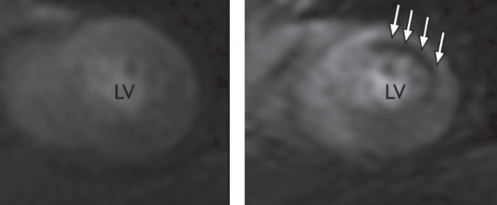
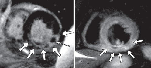
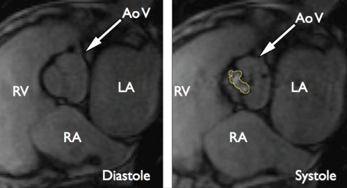
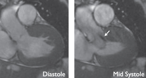
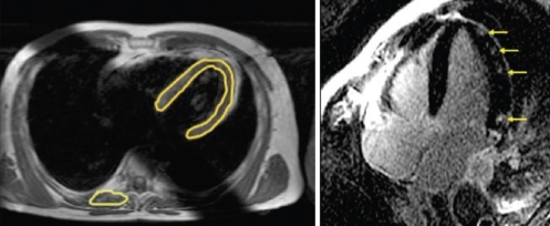
Starting as a research method little more than a decade ago, cardiovascular magnetic resonance (CMR) imaging has rapidly evolved to become a powerful diagnostic tool used in routine clinical cardiology. The contrast in CMR images is generated from protons in different chemical environments and, therefore, enables high-resolution imaging and specific tissue characterization in vivo, without the use of potentially harmful ionizing radiation.CMR imaging is used for the assessment of regional and global ventricular function, and to answer questions regarding anatomy. State-of-the-art CMR sequences allow for a wide range of tissue characterization approaches, including the identification and quantification of nonviable, edematous, inflamed, infiltrated or hypoperfused myocardium. These tissue changes are not only used to help identify the etiology of cardiomyopathies, but also allow for a better understanding of tissue pathology in vivo. CMR tissue characterization may also be used to stage a disease process; for example, elevated T2 signal is consistent with edema and helps differentiate acute from chronic myocardial injury, and the extent of myocardial fibrosis as imaged by contrast-enhanced CMR correlates with adverse patient outcome in ischemic and nonischemic cardiomyopathies.The current role of CMR imaging in clinical cardiology is reviewed, including coronary artery disease, congenital heart disease, nonischemic cardiomyopathies and valvular disease.
Utilisée comme mode de recherche il y a un peu plus de dix ans, l’imagerie cardiovasculaire par résonance magnétique (CRM) a rapidement évolué pour devenir un puissant outil diagnostique en cardiologie clinique régulière. Le contraste des images CRM est produit par les protons des divers environnements chimiques qui, par conséquent, procurent une imagerie à haute résolution et une caractérisation tissulaire spécifique in vivo, sans qu’il soit nécessaire d’utiliser le rayonnement ionisant au potentiel néfaste.
L’imagerie CRM permet d’évaluer la fonction ventriculaire régionale et globale et de répondre à des questions liées à l’anatomie. Les séquences CRM de pointe permettent toute une série d’approches de caractérisation tissulaire, y compris le dépistage et la quantification d’un myocarde non viable, œdémateux, enflammé, infiltré ou hyperperfusé. Ces changements tissulaires sont non seulement utilisés pour contribuer à repérer l’étiologie des myocardiopathies, mais permettent également de mieux comprendre la pathologie tissulaire in vivo. La caractérisation tissulaire CRM peut également permettre d’établir le stade du processus de la maladie. Par exemple, un signal T2 élevé est compatible avec un œdème et contribue à différencier une lésion myocardique aiguë d’une lésion chronique, et l’étendue de la fibrose myocardique perçue par IRM avec injection d’un agent de contraste est corrélée avec des issues négatives en cas de myocardiopathie ischémique ou non ischémique.
Le rôle actuel de l’imagerie CRM en cardiologie clinique est analysé, y compris la coronaropathie, la cardiopathie congénitale, les myocardiopathies non ischémiques et les affections valvulaires.
Figures
References
-
- Hendel RC, Patel MR, Kramer CM, et al. ACCF/ACR/SCCT/SCMR/ASNC/NASCI/SCAI/SIR 2006 appropriateness criteria for cardiac computed tomography and cardiac magnetic resonance imaging: A report of the American College of Cardiology Foundation Quality Strategic Directions Committee Appropriateness Criteria Working Group, American College of Radiology, Society of Cardiovascular Computed Tomography, Society for Cardiovascular Magnetic Resonance, American Society of Nuclear Cardiology, North American Society for Cardiac Imaging, Society for Cardiovascular Angiography and Interventions, and Society of Interventional Radiology. J Am Coll Cardiol. 2006;48:1475–97. - PubMed
-
- Beanlands RS, Chow BJ, Dick A, et al. CCS/CAR/CANM/CNCS/CanSCMR joint position statement on advanced noninvasive cardiac imaging using positron emission tomography, magnetic resonance imaging and multidetector computed tomographic angiography in the diagnosis and evaluation of ischemic heart disease – executive summary. Can J Cardiol. 2007;23:107–19. - PMC - PubMed
-
- Simonetti OP, Cook S. Technical aspects of pediatric CMR. J Cardiovasc Magn Reson. 2006;8:581–93. - PubMed
-
- Valente AM, Powell AJ. Clinical applications of cardiovascular magnetic resonance in congenital heart disease. Cardiol Clin. 2007;25:97–110. vi. - PubMed
Publication types
MeSH terms
LinkOut - more resources
Full Text Sources
Medical
