. 2010 Nov;19(11):1936-41.
doi: 10.1007/s00586-010-1478-7. Epub 2010 Jun 13.

Anterior approaches to the cervicothoracic junction: a study on the surgical accessibility of three different corridors based on the CT images

Affiliations

Anterior approaches to the cervicothoracic junction: a study on the surgical accessibility of three different corridors based on the CT images

Yi-Xing Huang et al. Eur Spine J. 2010 Nov.

Abstract

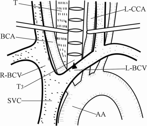
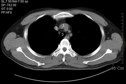
To determine the location of left brachiocephalic vein (BCV) and tracheal bifurcation (TB) relative to the vertebral levels, and to ascertain the accessibility of three different corridors (C1: between the esophagus and trachea medially and the carotid sheath laterally, C2: between the right BCV and the brachiocephalic artery, and C3: between the ascending aorta and superior vena cava) for preoperative planning. From August 2008 to April 2009, normal chest CT scans of 150 subjects ranging in age from 18 to 78 years were selected. According to our definition, of the 150 studies, 132 T2 vertebral bodies (VBs) could be accessed through C1 (88.0%), 100 T3 VBs could be reached through C2 (66.7%), and 110 T4 VBs could be exposed through C3 (73.3%). The results suggest that the surgical accessibility of three different corridors is different and we conclude that T2, T3, and T4 are, respectively, readily accessible through C1, C2, and C3.


Figures

Fig. 1
Fig. 1
The superior corridor (C1). AA ascending aorta, BCA brachiocephalic artery, L-BCV left brachiocephalic vein, L-CCA left common carotid artery, R-BCV right brachiocephalic vein, SVC superior vena cava, T trachea, T3 the third thoracic vertebral body, the black triangle represents the intersection between the superior margin of the LBCV and the mid-sagittal plane which determines the caudal accessibility of C1
Fig. 2
Fig. 2
The middle corridor (C2). AA ascending aorta, BCA brachiocephalic artery, L-BCV left brachiocephalic vein, L-CCA left common carotid artery, R-BCV right brachiocephalic vein, SVC superior vena cava, T4 the fourth thoracic vertebral body, the black triangle represents the superior border of the confluence of the left and right BCV which determines the caudal accessibility of C2
Fig. 3
Fig. 3
The inferior corridor (C3). AA ascending aorta, BCA brachiocephalic artery, L-BCV left brachiocephalic vein, L-CCA left common carotid artery, R-BCV right brachiocephalic vein, SVC superior vena cava, TB tracheal bifurcation, T5 the fifth thoracic vertebral body, the black triangle represents the superior border of the TB which determines the caudal accessibility of C3
Fig. 4
Fig. 4
A chest CT image (right) and its scout image (left) on the PACS workstation
Fig. 5
Fig. 5
The transverse plane of the intersection between the superior margin of the LBCV and the mid-sagittal plane. 1 left brachiocephalic vein, 2 right brachiocephalic vein, 3 trachea
Fig. 6
Fig. 6
The transverse plane of the superior border of the confluence of the left and right brachiocephalic veins. 1 left brachiocephalic vein, 2 right brachiocephalic vein, 3 trachea
Fig. 7
Fig. 7
The transverse plane of the superior border of the tracheal bifurcation. 1 tracheal bifurcation, 2 superior vena cava, 3 ascending aorta, 4 descending aorta

References

    1. An HS, Wise JJ, Xu R. Anatomy of the cervicothoracic junction: a study of cadaveric dissection, cryomicrotomy, and magnetic resonance imaging. J Spinal Disord. 1999;12:519–525. doi: 10.1097/00002517-199912000-00012. - DOI - PubMed
    1. Birch R, Bonney G, Marshall RW. A surgical approach to the cervicothoracic spine. J Bone Jt Surg Br. 1990;72:904–907. - PubMed
    1. Boockvar JA, Philips MF, Telfeian AE, et al. Results and risk factors for anterior cervicothoracic junction surgery. J Neurosurg. 2001;94:12–17. - PubMed
    1. Cauchoix J, Binet JP. Anterior surgical approaches to the spine. Ann R Coll Surg Engl. 1957;21:237–243. - PMC - PubMed
    1. Charles R, Govender S. Anterior approach to the upper thoracic vertebrae. J Bone Jt Surg Br. 1989;71:81–84. - PubMed

MeSH terms