. 2010 Oct;343(1-2):217-22.
doi: 10.1007/s11010-010-0516-4. Epub 2010 Jun 13.

Oxidized LDL up-regulates the ascorbic acid transporter SVCT2 in endothelial cells

Affiliations

Oxidized LDL up-regulates the ascorbic acid transporter SVCT2 in endothelial cells

James M May et al. Mol Cell Biochem. 2010 Oct.

Abstract

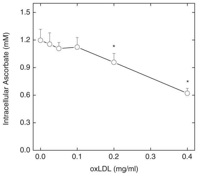
Endothelial dysfunction is an early manifestation of atherosclerosis caused in part by oxidized LDL (oxLDL). Since vitamin C, or ascorbic acid, prevents several aspects of endothelial dysfunction, the effects of oxLDL on oxidative stress and regulation of the ascorbate transporter, SVCT2, were studied in cultured EA.hy926 endothelial cells. Cells cultured for 18 h with 0.2 mg/ml oxLDL showed increased lipid peroxidation that was prevented by a single addition of 0.25 mM ascorbate at the beginning of the incubation. This protection caused a decrease in intracellular ascorbate, but no change in the cell content of GSH. In the absence of ascorbate, oxLDL increased SVCT2 protein and function during 18 h in culture. Although culture of the cells with ascorbate did not affect SVCT2 protein expression, the oxLDL-induced increase in SVCT2 protein expression was prevented by ascorbate. These results suggest that up-regulation of endothelial cell SVCT2 expression and function may help to maintain intracellular ascorbate during oxLDL-induced oxidative stress, and that ascorbate in turn can prevent this effect.


Figures

Figure 1
Figure 1
Oxidative stress induced by oxLDL and protection by ascorbate. EA.hy926 cells were cultured in the presence (circles) or absence (square) of 0.2 mg/ml oxLDL and the indicated ascorbate concentration. After 18 h, aliquots of the medium were removed for assay of malondialdehyde (Panel B) and the cells were rinsed twice in KRH and taken for assay of malondialdehyde (Panel A) and GSH (Panel C). Results are shown from 4 or more experiments, with an “*” indicating p < 0.05 compared to time zero.
Figure 2
Figure 2
OxLDL decreases intracellular ascorbate. Cells were cultured with 0.25 mM ascorbate and the indicated concentration of oxLDL. After 18 h, the cells were rinsed 3 times in KRH and taken for assay of intracellular ascorbate. Results are shown from 5 experiments, with “*” indicating p < 0.05 compared to cells not treated with oxLDL.
Figure 3
Figure 3
OxLDL increases SVCT2 message and protein. EA.hy926 cells were cultured for 18 h with the indicated concentrations of oxLDL then taken for assay of SVCT2 protein by immunoblotting. Panel A shows a representative immunoblot of the SVCT2, using β-actin as a control for gel loading. Panel B shows the densitometry from 5 assays of SVCT2 protein expression, normalized to β-actin and expressed as mean + SD in standardized units of density (circles). The single square shows data from 3 experiments for the indicated concentration of non-oxidized LDL. Panel C shows oxLDL effects on radiolabeled ascorbate transport. An asterisk (*) indicates p < 0.05 compared to the sample not treated with oxLDL.
Figure 4
Figure 4
Ascorbate effects on oxLDL-induced increases SVCT2. EA.hy926 cells were cultured for 18 h with the indicated concentrations of oxLDL and ascorbate, and then taken for assay of SVCT2 expression by immunoblotting. Panel A. shows a representative immunoblot of the SVCT2, with β-actin as a control for gel loading. Panel B shows the densitometry from 5 assays of SVCT2 protein expression, normalized to β-actin and expressed as mean + SD in arbitrary density units. Bars not sharing the same letters are different from one another at p < 0.05.

Similar articles

Cited by

References

    1. Steinberg D, Parthasarathy S, Carew TE, et al. Modifications of low-density lipoprotein that increase its atherogenicity. N Engl J Med. 1989;320:915–924. - PubMed
    1. Steinbrecher UP, Parthasarathy S, Leake DS, et al. Modification of low density lipoprotein by endothelial cells involves lipid peroxidation and degradation of low density lipoprotein phospholipids. Proc Natl Acad Sci USA. 1984;81:3883–3887. - PMC - PubMed
    1. Steinbrecher UP. Role of superoxide in endothelial-cell modification of low-density lipoproteins. Biochim Biophys Acta. 1988;959:20–30. - PubMed
    1. Schmitt A, Salvayre R, Delchambre J, et al. Prevention by α-tocopherol and rutin of glutathione and ATP depletion induced by oxidized LDL in cultured endothelial cells. Br J Pharmacol. 1995;116:1985–1990. - PMC - PubMed
    1. Martin A, Frei B. Both intracellular and extracellular vitamin C inhibit atherogenic modification of LDL by human vascular endothelial cells. Arterioscler Thromb Vasc Biol. 1997;17:1583–1590. - PubMed

Publication types

MeSH terms

Substances