. 2010 Oct;58(13):1553-69.
doi: 10.1002/glia.21029.

Origin, maturation, and astroglial transformation of secondary radial glial cells in the developing dentate gyrus

Affiliations

Origin, maturation, and astroglial transformation of secondary radial glial cells in the developing dentate gyrus

Bianka Brunne et al. Glia. 2010 Oct.

Abstract

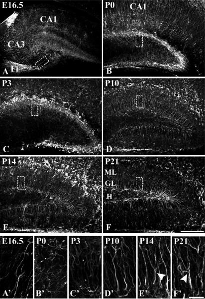
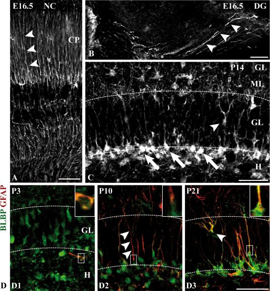
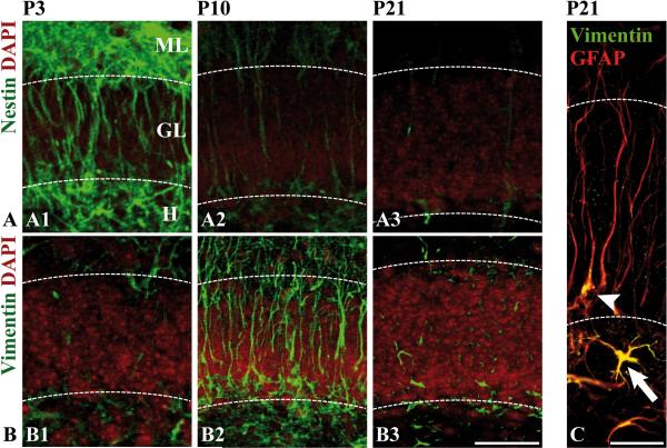
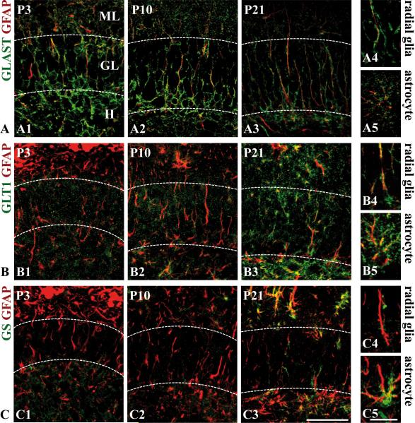
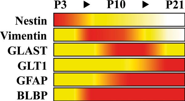
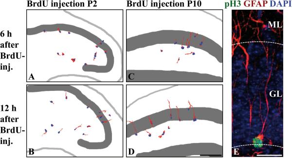
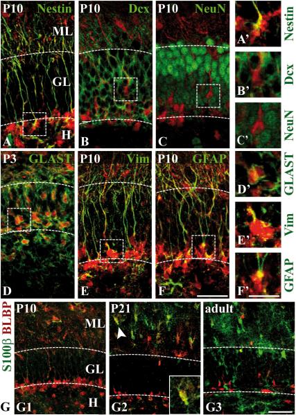
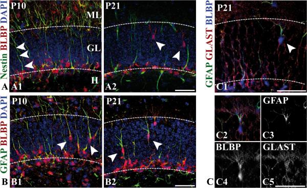
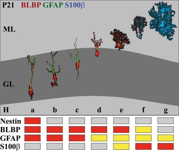
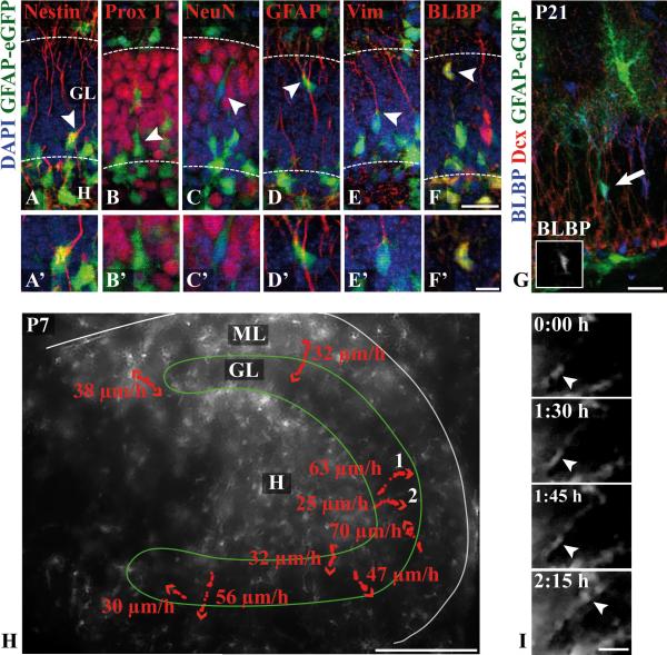
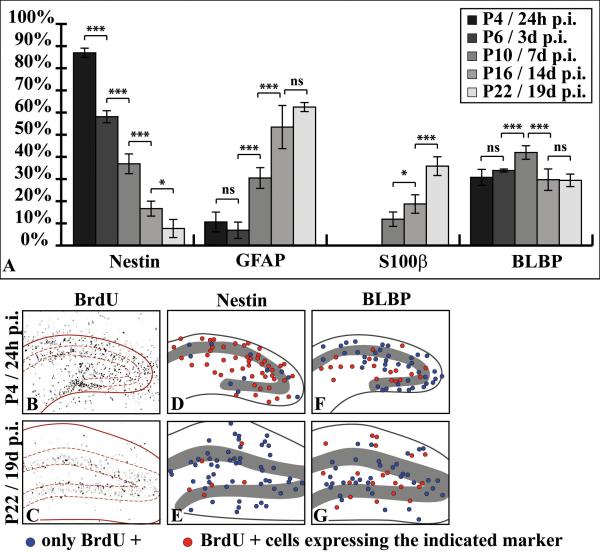
The dentate gyrus is a brain region where neurons are continuously born throughout life. In the adult, the role of its radial glia in neurogenesis has attracted much attention over the past years; however, little is known about the generation and differentiation of glial cells and their relationship to radial glia during the ontogenetic development of this brain structure. Here, we combine immunohistochemical phenotyping using antibodies against glial marker proteins with BrdU birthdating to characterize the development of the secondary radial glial scaffold in the dentate gyrus and its potential to differentiate into astrocytes. We demonstrate that the expression of brain lipid-binding protein, GLAST, and glial fibrillary acidic protein (GFAP) characterizes immature differentiating cells confined to an astrocytic fate in the early postnatal dentate gyrus. On the basis of our studies, we propose a model where immature astrocytes migrate radially through the granule cell layer to adopt their final positions in the molecular layer of the dentate gyrus. Time-lapse imaging of acute hippocampal slices from hGFAP-eGFP transgenic mice provides direct evidence for such a migration mode of differentiating astroglial cells in the developing dentate gyrus.


Figures

Figure 1
Figure 1. Development of the radial glial scaffold in the murine dentate gyrus
Immunohistochemical visualization of radial glial fibers with a rabbit polyclonal GFAP antibody in coronal hippocampal sections of the indicated developmental stages. The boxed regions are shown at higher magnification in (A’-F’), which are extended focus images from stacks of 10 serial z-sections of 1 μm. (A,A’) At E16.5, fibers of the primary radial glial scaffold extend from the proliferative zone above the prospective fimbria (Fi) towards the developing suprapyramidal blade of the dentate gyrus. (B,B’) At P0, GFAP-positive fibers are short and lack a radial arrangement. (C,C’) Emergence of a secondary radial glial scaffold at P3: radial fibers extend from the hilar region towards the developing molecular layer. (D-D’, E-E’) During the second postnatal week, the secondary radial glial scaffold has fully evolved. Regularly arranged radial fibers traverse the supra- and infrapyramidal blades of the granular layer. (F,F’) Secondary scaffold at P21. Note the occurence of branched glial fibers at later stages (arrowheads in E’,F’). Astrocytic cells in the outer molecular layer and hilus are also immunolabeled at late stages. Scale bars: 200 μm (A-F), 25 μm (A’-F’). GL, granular layer; H, hilus; ML, molecular layer.
Figure 2
Figure 2. BLBP as radial glial marker in the developing dentate gyrus
(A) Neocortex (NC): BLBP immunolabels radial glial fibers (arrowheads) in the developing cortical plate (CP). (B) Dentate gyrus (DG): At E16.5, strong BLBP immunoreactivity is detected in the processes of primordial radial glial cells (arrowheads) in the dentate gyrus (extended focus image from a stack of 10 serial z-sections of 0.55 μm). (C) At P14, BLBP immunoreactivity is primarily found in the somata (arrows) of secondary radial glia in the subgranular zone (SGZ). Non-radial BLBP-positive cell bodies are also localized within the GL (arrowhead) and in the ML. (D) Hippocampal sections of the developmental stages P3, P10 and P21 were immunostained with an antibody against BLBP (green) and a monoclonal antibody against GFAP (red). The boxed images are magnifications of the cells within the dotted frames and demonstrate colocalization of BLBP and GFAP. The radial processes (arrowheads in D2) are only weakly BLBP-immunoreactive. Note the branched GFAP- and BLBP-positive cell within the granular layer at P21 (arrowhead, D3). Scale bars: 50 μm. GL, granular layer; H, hilus; ML, molecular layer.
Figure 3
Figure 3. Intermediate filament protein expression in secondary radial glia
Hippocampal sections of the developmental stages P3, P10 and P21 were immunostained with antibodies against nestin (A1-A3) or vimentin (B1-B3). Nuclei were counterstained with DAPI (red) to visualize the granule cell layer (GL). Nestin (green) labels secondary radial glial cells at early and intermediate stages (A1-A2), whereas vimentin (green) is a predominant marker at intermediate stages (B2). (C) Detection of vimentin in hilar astrocytes (arrow) and radial glia (arrowhead) at P21. Coimmunostaining with the polyclonal GFAP antibody (red) was used to visualize the radial processes. - Scale bars: 50 μm (C: 25 μm).
Figure 4
Figure 4. Expression profile of glial markers related to glutamate metabolism in secondary radial glia
Hippocampal sections of the developmental stages P3, P10 and P21 were immunostained with antibodies against GLAST (A1-A5), GLT1 (B1-B5), and glutamine synthetase (GS) (C1-C5). To visualize radial processes, the sections were counterstained with the polyclonal GFAP antibody (red). GLAST immunoreactivity in radial glia was already detected at P3 (A1) and strongest at P10 (A2). (A4) A GLAST-positive radial glial process at P21. Weak GLT1-immunoreactivity of radial glial cells was only seen at late stages (B3-4), whereas no glutamine synthetase expression was detectable in secondary radial glia (C1-C4). GLAST (A5), GLT1 (B5) and glutamine synthetase (C5) expression in astrocytes at P21. Scale bars: 50 μm (A1-3,B1-3, C1-3), 10 μm (A4-5,B4-5,C4-5). GL, granular layer; H, hilus; ML, molecular layer.
Figure 5
Figure 5. Expression profile of glial marker proteins in secondary radial glia
The relative immunostaining of different glial marker proteins during dentate gyrus development in the secondary radial scaffold is represented by a colour code: White, barely detectable; yellow, moderate immunostaining of many cells or strong labeling of scattered individual cells; red, strong immunoreactivity of many cells. S100-beta and glutamine synthetase are not included; no significant expression of these markers was observed in secondary radial glia.
Figure 6
Figure 6. Assembly and proliferative capacity of the secondary radial glial scaffold
(A-D) Proliferating cells in the developing dentate gyrus were birthdated with BrdU (blue) at P2 (A-B) or P10 (C-D) and immunostained for nestin. Processes were reconstructed based on their nestin immunoreactivity (red) using a Photoshop selection tool. (A) 6hr after BrdU-injection, labeled nestin-positive proliferating cells do not yet possess radial processes. (B) 12hr later, the majority of BrdU-labeled cells has outgrown a nestin-positive radial process, indicating that secondary radial glial cells are derived from proliferating cells in the hilus at P2. (C-D) 6 or 12hr after BrdU injection at P10, most of the labeled cells display a nestin-positive radial process, identifying proliferating cells at this stage as belonging to the secondary radial glial scaffold. (E) A pH3-positive (green), mitotic secondary radial glial cell at P14, characterized by a long unbranched GFAP-positive process (red) traversing the GL. The soma is localized in the subgranular zone at the border between hilus and GL. Scale bars: 200 μm (A-D), 25 μm (E). GL, granule cell layer; ML, molecular layer.
Figure 7
Figure 7. Coexpression of BLBP with astroglial differentiation markers in the developing dentate gyrus
(A-F) Coronal sections of the suprapyramidal granule cell layer at P10 (P3 in D) were coimmunostained with antibodies against BLBP (red) and the indicated marker (green). The boxed regions are shown at higher magnification in (A’-F’). (A) A strong colocalization of BLBP and the precursor cell marker nestin is detected in radial glial cells in the subgranular zone. (B-C) No colocalization of BLBP with the neuronal markers doublecortin (Dcx) (B) or NeuN (C) can be observed. (D) Double-labeling of BLBP and GLAST is most prominent at P3. (E-F) Colocalization of BLBP with vimentin (Vim) and GFAP (immunolabeled with the mouse monoclonal antibody) in the secondary radial glial scaffold peaks in the second postnatal week. (G1-G3) Coimmunostaining of BLBP (red) and S100-beta (green) at P10, P21 and in adult mice. Occasional colocalization of both markers in astroglial cells of the inner ML is observed at P21 (G2). The inset shows one of the double-labeled cells (arrowhead) at higher magnification. Coexpression is neither seen at P10, where almost no S100-beta can be observed (G1), nor in adult mice, where BLBP-positive cells in the ML are rarely detected (G3). Note the increase in S100-beta expression over time. Scale bars: 50 μm (A-F, G1-G3), 25 μm (A’-F’, G2’,G2’’). GL, granule cell layer; ML, molecular layer.
Figure 8
Figure 8. Immunohistochemical characterization of transforming secondary radial glia
(A1-A2) At P10 (A1), BLBP (red) and nestin (green) are colocalized in secondary radial glial cells, which extend unbranched radial processes through the granule cell layer. At P21 (A2), BLBP-positive cells whose somata are located within the granular layer extend branched processes, which are negative for nestin (arrowheads). Nuclei were counterstained with DAPI. (B1-B2), Colocalization of BLBP (red) and GFAP (green, monoclonal antibody) at P10 and P21. At P10 (B1), somata of double-immunolabeled radial glial cells with unbranched processes are localized within the granule cell layer (arrowheads). At P21 (B2), BLBP-positive cells whose somata are located within the granular layer extend branched GFAP-immunoreactive processes (arrowheads). Nuclei were counterstained with DAPI. (C1-C5) Triple-immunostaining of a hippocampal section at P21 with antibodies against BLBP (blue), GLAST (red) and GFAP (green, monoclonal antibody). (C2) Single-channel views of the triple-labeled cell (arrowhead in C1) are shown in C3-C5. Note the membranous punctuate GLAST immunoractivity (C5). - Scale bars: 50 μm.
Figure 9
Figure 9. A model of radial glial migration and astrocytic transformation in the developing dentate gyrus
(a) The somata of secondary radial glial cells in the developing dentate gyrus are located in the subgranular zone and extend long radial processes that traverse the granular layer (GL). At this stage secondary radial glia with progenitor cell properties express nestin, BLBP (red) and GFAP (green). Astroglial differentiation is characterized by downregulation of nestin expression (b) and morphological changes: the BLBP-positive soma translocates from the subgranular zone into the granule cell layer, while the GFAP-positive process starts to ramify within the granular layer. Increasing branching of the processes occurs while the cells translocate through the GL towards the molecular layer (ML) (b-d). Immature astrocytes at the border to and within the ML start to express S100-beta and gradually lose BLBP expression (d-f). Mature astrocytes in the molecular layer are characterized by the expression of S100-beta and in some cases of GFAP (g). – All of the displayed stages of radial glial transformation were taken from sections of 21 day-old mice using a Photoshop selection tool. The grading of the immunstaining was colour-coded as described in Figure 5.
Figure 10
Figure 10. In vivo imaging of astroglial migration in the developing murine dentate gyrus
(A-F) Immunohistochemical characterization of GFP-fluorescent cells in the dentate gyrus of P7 hGFAP-eGFP transgenic mice. Coronal sections were immunostained with antibodies against the indicated markers (red) and counterstained with DAPI (blue). Colocalization of GFP fluorescence (green) with nestin (A) and astroglial (D-F) but not with neuronal markers (B-C) is observed (arrowheads). Magnifications of the marked cells are shown in (A’-F’). (G) Colocalization of BLBP (blue) and GFP fluorescence in a translocating glial cell (arrow) whose soma is located in the middle of the granular layer (inset in G to show single BLBP staining). No colocalization of GFP fluorescence and Dcx (red) is observed. (H) Fluorescent live microscopy of a hippocampal slice culture prepared from a P7 hGFAP-eGFP transgenic mouse. The positions of different cells were monitored every 15 min and marked by red dots. The arrowheads indicate the direction of migrating cells. The maximal cell speed (μm/hr) of the highlighted cells are indicated. A total of 19 cells from two different preparations were analyzed. (I) Fluorescent time-lapse images of cell 1 (arrowhead: soma). Scale bars: 25 μm (A-F,I), 10 μm (A’-F’), 50 μm (G), 200 μm (H). Dcx, doublecortin; GL, granular layer; H, hilus; ML, molecular layer; Vim, vimentin.
Figure 11
Figure 11. Immunohistochemical profiling of P3-birthdated progenitor cells at various stages of dentate gyrus development
(A) The diagram shows the percentage (mean ±SD) of BrdU-labeled cells expressing nestin, GFAP, S100-beta or BLBP at the indicated time points after BrdU injection at P3 (n=180 cells). Differentiating cells quickly downregulate nestin, while GFAP and S100-beta are upregulated. BLBP expression did not change much during the period investigated. (B-C) Proliferating cells in the dentate gyrus were BrdU-labeled at P3 and tracked 24hr (P4) and 19 days (P22) post injection (p.i.). (B) 24hr p.i., BrdU-labeled cells are distributed over the entire area of the developing hippocampus. (C) At P22, the cells that were labeled at P3 are primarily found in the subgranular zone and in the granular layer. (D-G) To determine the redistribution of labeled cells during development, 20 BrdU-positive cells in the hilus, granular and molecular layer were arbitrarily chosen and tested for expression of nestin (D-E) or BLBP (F-G), respectively. Blue circles indicate BrdU-only positive cells, red circles represent BrdU-positive cells that express the respective marker. Most of the BrdU-labeled cells are nestin-positive at P4 (D), whereas double-labeled cells at P22 are rare and mostly confined to the subgranular zone (E). BLBP-positive BrdU-positive cells are scattered over the dentate area at P4 (F) and redistribute to the hilus, molecular layer and subgranular zone at P22 (G).

References

    1. Altman J, Bayer SA. Migration and distribution of two populations of hippocampal granule cell precursors during the perinatal and postnatal periods. J Comp Neurol. 1990a;301(3):365–81. - PubMed
    1. Altman J, Bayer SA. Mosaic organization of the hippocampal neuroepithelium and the multiple germinal sources of dentate granule cells. J Comp Neurol. 1990b;301(3):325–42. - PubMed
    1. Alves JA, Barone P, Engelender S, Froes MM, Menezes JR. Initial stages of radial glia astrocytic transformation in the early postnatal anterior subventricular zone. J Neurobiol. 2002;52(3):251–65. - PubMed
    1. Anthony TE, Heintz N. Genetic lineage tracing defines distinct neurogenic and gliogenic stages of ventral telencephalic radial glial development. Neural Develop. 2008;3(1):30. - PMC - PubMed
    1. Barry D, McDermott K. Differentiation of radial glia from radial precursor cells and transformation into astrocytes in the developing rat spinal cord. Glia. 2005;50(3):187–97. - PubMed

Publication types

MeSH terms

Substances