. 2010 Sep 15;116(18):4420-32.
doi: 10.1002/cncr.25322.

Adenovirus-mediated expression of truncated E2F-1 suppresses tumor growth in vitro and in vivo

Affiliations

Adenovirus-mediated expression of truncated E2F-1 suppresses tumor growth in vitro and in vivo

Jorge G Gomez-Gutierrez et al. Cancer. .

Expression of concern in

  • Expression of concern.
    [No authors listed] [No authors listed] Cancer. 2024 Sep 1;130(17):3047. doi: 10.1002/cncr.35435. Epub 2024 Jun 26. Cancer. 2024. PMID: 38922873 No abstract available.

Abstract

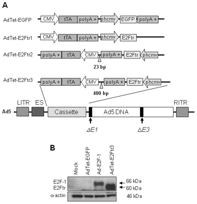
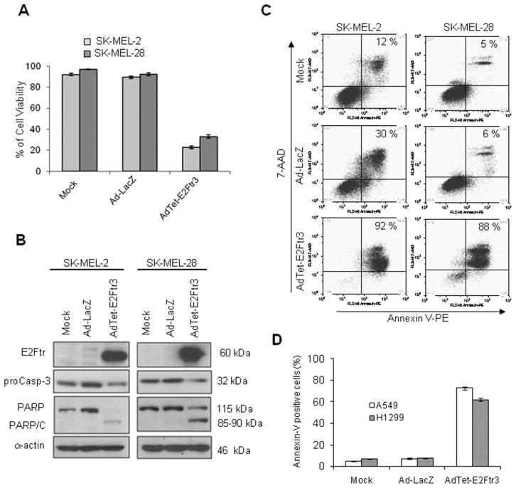
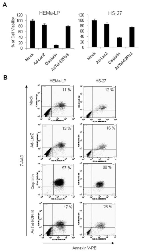
Background: Adenovirus (Ad)-mediated E2F-1 gene transfer induces apoptosis in cancer cells in vitro and in vivo, but clinical application of E2F-1 in cancer gene therapy remains controversial because of the oncogenic potential of E2F-1. This barrier can be circumvented by using the truncated form of the E2F-1 gene (E2Ftr) (amino acids 1 through 375), which lacks the E2F-1 transactivation domain and cell cycle-promoting effects.

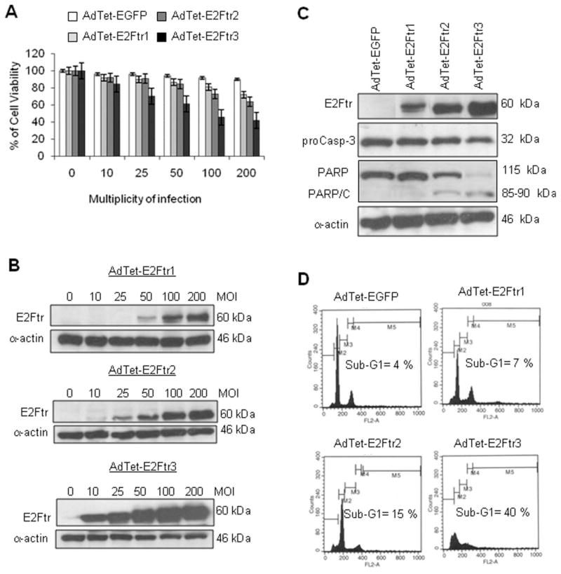
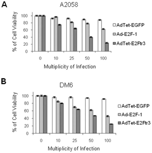
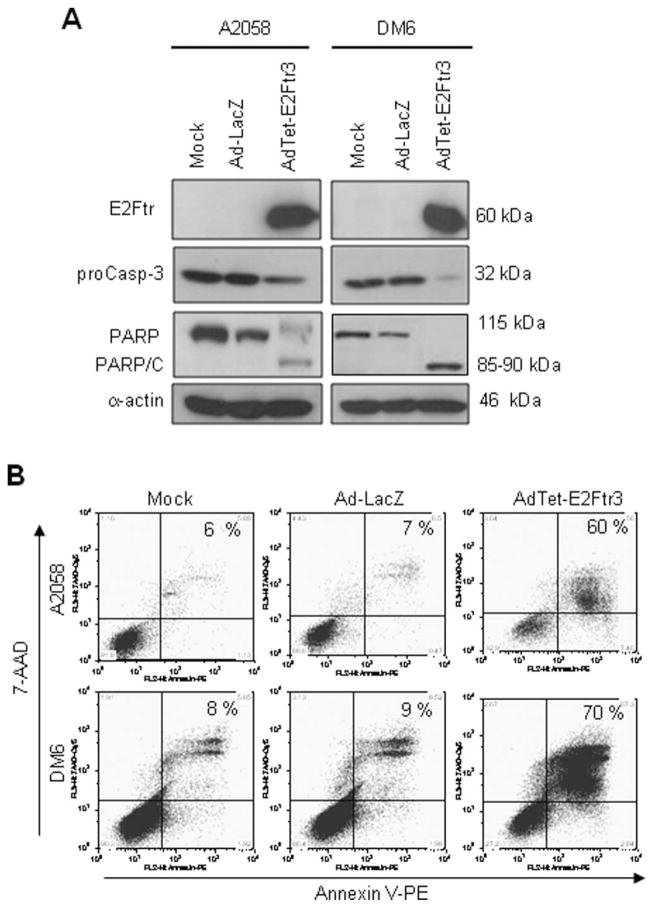
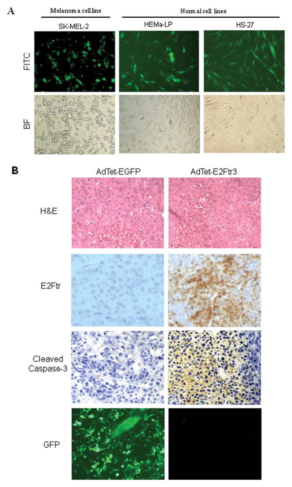
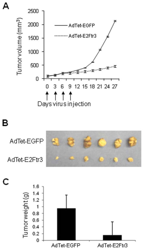
Methods: The authors constructed 3 adenoviral vectors that expressed E2Ftr under regulation of the tetracycline (Tet)-off system (AdTet-E2Ftr1, AdTet-E2Ftr2, and AdTet-E2Ftr3). These vectors were compared for E2Ftr expression and apoptosis induction in cancer cells and normal cells. E2Ftr antitumor activity in vivo also was assessed in a melanoma xenograft model.

Results: One of the 3 vectors, AdTet-E2Ftr3, had the highest E2Ftr protein expression levels, which were correlated with the greatest induction of apoptosis and inhibition of cancer cell growth. E2Ftr induced apoptosis in a variety of cancer cell lines independent of p53 status with little cytotoxicity in normal cell lines. In a mouse melanoma xenograft model, AdTet-E2Ftr3 exhibited an approximately 80% decrease in tumor size compared with controls in vivo.

Conclusions: The current results indicated that AdTet-E2Ftr3 is a novel anticancer agent that has significant therapeutic activity in vitro and in vivo.


Figures

Figure 1
Figure 1
This is a schematic diagram of 4 single, bicistronic adenovirus (Ad) vectors and confirms deletion of the E2F-1 transactivation domain. (A) The cytomegalovirus (CMV) promoter drives expression of the tetracycline (Tet)-controlled transactivator (tTA). The vector that expresses the reporter-enhanced green fluorescent protein (EGFP) or the truncated form of the E2F-1 gene (E2Ftr) is under the control of a synthetic minimal promoter composed of a Tet-responsive element (TRE) and a CMV mini-promoter (phcmv), which is silent unless activated by tTA. In the absence of Tet or doxycycline, tTA binds to phcmv and triggers the expression of EGFP or E2Ftr. When Tet is added to the medium, tTA is bound by Tet and is unable to bind to phcmv and activate the expression of EGFP or E2Ftr. The left and right inverted terminal repeat sequences (LITR and RITR, respectively), encapsidation signal (ES), and E1/E3-deleted genes are shown in the Ad structure. polyA indicates RNA with the addition of multiple adenosine monophosphates. (B) SK-MEL-2 cells were uninfected (Mock) or were infected with AdTet-EGFP, Ad-E2F-1, or AdTet-E2Ftr3 at a multiplicity of infection of 100; and, 36 hours postinfection, monoclonal antibody antihuman E2F-1 was used to detect both E2F-1 and E2Ftr expression. α-Actin was used to demonstrate equal loading for each lane.
Figure 2
Figure 2
Three adenovirus (Ad) vectors that expressed the truncated form of the E2F-1 gene (E2Ftr) were evaluated for their ability to induce melanoma cell killing and apoptosis. SK-MEL-2 cells were infected with Ad vectors that expressed E2Ftr under regulation of the tetracycline (Tet)-off system (AdTet-E2Ftr1, AdTet-E2Ftr2, and AdTet-E2Ftr3 or AdTet that expressed the reporter-enhanced green fluorescent protein [AdTet-EGFP]) at multiplicities of infection (MOIs) of 0, 10, 25, 50, 100, or 200. Three days after infection, cells were used for 3-(4,5-dimethylthiazol-2-yl)-2,5-diphenyltetrazolium (MTT) and Western blot assays. (A) The MTT assay was used to determine cell survival. Each point represents the mean of 3 independent experiments (±standard deviation [bars]). (B) Monoclonal antibody (MoAb) antihuman E2F-1 was used to detect E2Ftr. SK-MEL-2 cells were infected with the vectors mentioned above at an MOI of 100. Three days after infection, the cells were used for a Western blot assay and cell cycle analysis. (C) MoAb antihuman poly (ADP-ribose) polymerase (PARP) was used to detect PARP, and MoAb antihuman caspase-3/CPP3 was used to detect caspase-3 (proCasp-3). α-Actin was used to demonstrate equal loading for each lane. PARP/C indicates PARP cleavage. (D) For cell cycle analysis, cells were stained with propidium iodide and analyzed as describe in the text (see Materials and Methods).
Figure 3
Figure 3
An E2F-1 adenovirus (Ad-E2F-1) vector was compared with an Ad vector that expressed the truncated form of the E2F-1 gene (E2Ftr) under regulation of the tetracycline (Tet)-off system (AdTet-E2Ftr3) in an assay that inhibited cell proliferation. (A) A2058 cells and (B) DM6 cells were infected with AdTet-expressing enhanced green fluorescent protein (AdTet-EGFP), Ad-E2F-1, or AdTet-E2Ftr3 at multiplicities of infection of 0, 10, 25, 50, or 100. Seventy-two hours after infection, the cells were used for a 3-(4,5-dimethylthiazol-2-yl)-2,5-diphenyltetrazolium (MTT) assay to determine cell survival. Each point represents the mean of 3 independent experiments (±standard deviation [bars]).
Figure 4
Figure 4
The induction of apoptosis is illustrated by the truncated form of the E2F-1 gene (E2Ftr) in melanoma cells that were used in tumor xenograft models. A2058 cells and DM6 cells were uninfected (Mock) or were infected with a recombinant Ad serotype 5 vector encoding the Escherichia coli β-galactosidase (lacZ) gene controlled by the human cytomegalovirus (CMV) promoter (Ad5CMV-LacZ [Ad-LacZ]) or with an Ad vector that expressed E2Ftr under regulation of the tetracycline (Tet)-off system (AdTet-E2Ftr3) at a multiplicity of infection of 100. Seventy-two hours after infection, the cells were used for Western blot assays or Annexin V staining. (A) Monoclonal antibody (MoAb) antihuman E2F-1 was used to detect E2Ftr, antihuman poly (ADP-ribose) polymerase (PARP) was used to detect PARP, and MoAb antihuman caspase-3/CPP3 was used to detect caspase-3 (proCasp-3). α-Actin was used to demonstrate equal loading for each lane. PARP/C indicates PARP cleavage. (B) To determine the percentage of apoptosis, cells were stained with Annexin V-phycoerythrin (PE) and were analyzed as describe in the text (see Materials and Methods). 7-AAD indicates 7-aminoactinomycin D.
Figure 5
Figure 5
The role of p53 status on apoptosis induced by the truncated form of the E2F-1 gene (E2Ftr) is illustrated. SK-MEL-2 (mutant p53) cells and SK-MEL-28 (wild type [wt] p53) cells were uninfected (Mock) or were infected with either a recombinant adenovirus (Ad) serotype 5 vector encoding the Escherichia coli β-galactosidase (lacZ) gene controlled by the human cytomegalovirus (CMV) promoter (Ad5CMV-LacZ [Ad-LacZ]) or with an Ad vector that expressed E2Ftr under regulation of the tetracycline (Tet)-off system (AdTet-E2Ftr3) at a multiplicity of infection of 100. Seventy-two hours after infection, the cells were used for a 3-(4,5-dimethylthiazol-2-yl)-2,5-diphenyltetrazolium (MTT) and Western blot analyses or for Annexin V staining. (A) An MTT assay was used to determine cell survival. Each point represents the mean of 3 independent experiments (±standard deviation [bars]). (B) Monoclonal antibody (MoAb) antihuman E2F-1 was used to detect E2Ftr, MoAb antihuman poly (ADP-ribose) polymerase (PARP) was used to detect PARP, and MoAb antihuman caspase-3/CPP32 was used to detect caspase-3 (proCasp-3). α-Actin was used to demonstrate equal loading for each lane. PARP/C indicates PARP cleavage. (C) For Annexin V-phycoerythrin (PE) staining, cells were stained and analyzed as described in the text (see Materials and Methods). 7-AAD indicates 7-aminoactinomycin D. (D) A549 (wt p53) and H1299 (partial deletion of p53) lung cancer cells were treated as described above and in an apoptosis assay.
Figure 6
Figure 6
The cytotoxic effect of transduction in normal cell lines of an adenovirus (Ad) vector that expressed the truncated form of the E2F-1 gene (E2Ftr) under regulation of the tetracycline (Tet)-off system (AdTet-E2Ftr3) is illustrated. Normal HEMa-LP cells and HS-27 cells were uninfected, or infected with a recombinant Ad serotype 5 vector encoding the Escherichia coli β-galactosidase (lacZ) gene controlled by the human cytomegalovirus (CMV) promoter (Ad5CMV-LacZ [Ad-LacZ]) or with AdTet-E2Ftr3 at a multiplicity of infection of 100, or were treated with cisplatin 25 μM. Seventy-two hours after treatments, cell viability or apoptosis was analyzed by an 3-(4,5-dimethylthiazol-2-yl)-2,5-diphenyltetrazolium (MTT) assay or Annexin V staining, respectively. (A) For MTT, each point represents the mean of 3 independent experiments (±standard deviation [bars]). (B) For Annexin V staining, cells were stained and analyzed as describe in the text (see Materials and Methods). 7-AAD indicates 7-aminoactinomycin D; PE, phycoerythrin.
Figure 7
Figure 7
Normal cell transduction efficiency and tumor immunohistochemistry were evaluated. (A) SK-MEL-2, HEMa-LP, and HS-27 cells were transduced with an adenovirus (Ad) vector under regulation of the tetracycline (Tet)-off system (AdTet) that expressed enhanced green fluorescent protein (AdTet-EGFP) at a multiplicity of infection of 100. After 24 hours, the cells were examined for EGFP expression under fluorescence microscopy. Photomicrographs were taken with Kodak MDS 290 software at ×20 magnification. FITC indicates fluorescein isothiocyanate; BF, bright field. (B) Immunohistochemistry of melanoma tumors is shown 24 hours after the last treatment with AdTet-EGFP or with the AdTet vector that expressed the truncated form of the E2F-1 gene (E2Ftr) (AdTet-E2Ftr3) (hematoxylin and eosin [H&E] staining; original magnification, ×20). EGFP expression was observed with fluorescence microscopy. Monoclonal antibody antihuman E2F-1 was used to detect E2Ftr expression, and polyclonal antihuman cleaved caspase-3 (Asp175; 5A1E) was used to confirm apoptosis. GFP indicates green fluorescent protein.
Figure 8
Figure 8
In vivo antitumor effects of treatment with an adenovirus (Ad) vector that expressed the truncated form of the E2F-1 gene (E2Ftr) are illustrated. (A) After subcutaneous administration of 5 × 10⁶ A2058 cells, palpable tumors were randomized to receive a local injection of either an Ad that expressed E2Ftr under regulation of the tetracycline (Tet)-off system (AdTet) with enhanced green fluorescent protein (AdTet-EGFP) or the vector AdTet-E2Ftr3 on Days 0, 3, 6, and 9 (vertical arrows). The total viral dose was 4 × 2×1 × 109 plaque forming units. Tumor volume (V) was plotted against time and was determined by the equation V = (L × W2)/2, where L is the length, and W is the width of the tumor. (B) On Day 27, tumors were excised and photographed: The relative mass of tumors is shown. (C) Tumors also were weighed on Day 27. The results are expressed as the mean from each treatment group (±standard error [bars]). The differences in tumor size or weight in animals that were treated with AdTet-E2Ftr3 were statistically significant by the end of the experiments compared with controls (Day 27; P < .05).

Similar articles

Cited by

References

    1. Dong YB, Yang HL, Elliott MJ, et al. Adenovirus-mediated E2F-1 gene transfer efficiently induces apoptosis in melanoma cells. Cancer. 1999;86:2021–2033. - PubMed
    1. Dong YB, Yang HL, Elliott MJ, McMasters KM. Adenovirus-mediated E2F-1 gene transfer sensitizes melanoma cells to apoptosis induced by topoisomerase II inhibitors. Cancer Res. 2002;62:1776–1783. - PubMed
    1. Elliott MJ, Dong YB, Yang H, McMasters KM. E2F-1 up-regulates c-Myc and p14(ARF) and induces apoptosis in colon cancer cells. Clin Cancer Res. 2001;7:3590–3597. - PubMed
    1. Vorburger SA, Hetrakul N, Xia W, et al. Gene therapy with E2F-1 up-regulates the protein kinase PKR and inhibits growth of leiomyosarcoma in vivo. Mol Cancer Ther. 2005;4:1710–1716. - PubMed
    1. Itoshima T, Fujiwara T, Waku T, et al. Induction of apoptosis in human esophageal cancer cells by sequential transfer of the wild-type p53 and E2F-1 genes: involvement of p53 accumulation via ARF-mediated MDM2 down-regulation. Clin Cancer Res. 2000;6:2851–2859. - PubMed

Publication types

Substances