

The fatal attraction between pro-prion and filamin A: prion as a marker in human cancers
- PMID: 20550479
- PMCID: PMC2925173
- DOI: 10.2217/bmm.10.14
The fatal attraction between pro-prion and filamin A: prion as a marker in human cancers
Abstract
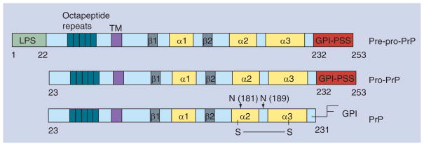
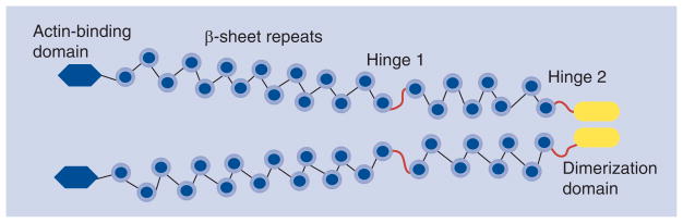
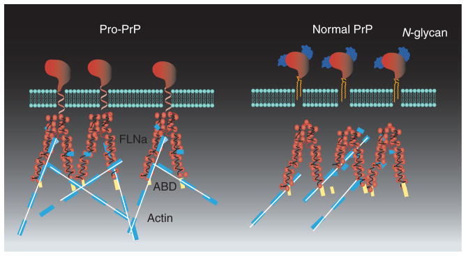
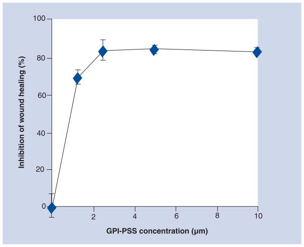
Pancreatic cancer is the fourth leading cancer causing deaths in the USA, with more than 30,000 deaths per year. The overall median survival for all pancreatic cancer is 6 months and the 5-year survival rate is less than 10%. This dismal outcome reflects the inefficacy of the chemotherapeutic agents, as well as the lack of an early diagnostic marker. A protein known as prion (PrP) is expressed in human pancreatic cancer cell lines. However, in these cell lines, the PrP is incompletely processed and exists as pro-PrP. The pro-PrP binds to a molecule inside the cell, filamin A (FLNa), which is an integrator of cell signaling and mechanics. The binding of pro-PrP to FLNa disrupts the normal functions of FLNa, altering the cell's cytoskeleton and signal transduction machineries. As a result, the tumor cells grow more aggressively. Approximately 40% of patients with pancreatic cancer express PrP in their cancer. These patients have significantly shorter survival compared with patients whose pancreatic cancers lack PrP. Therefore, expression of pro-PrP and its binding to FLNa provide a growth advantage to pancreatic cancers. In this article, we discuss the following points: the biology of PrP, the consequences of binding of pro-PrP to FLNa in pancreatic cancer, the detection of pro-PrP in other cancers, the potential of using pro-PrP as a diagnostic marker, and prevention of the binding between pro-PrP and FLNa as a target for therapeutic intervention in cancers.
Figures
Similar articles
-
Binding of pro-prion to filamin A: by design or an unfortunate blunder.Oncogene. 2010 Sep 30;29(39):5329-45. doi: 10.1038/onc.2010.307. Epub 2010 Aug 9. Oncogene. 2010. PMID: 20697352 Free PMC article. Review.
-
Binding of pro-prion to filamin A disrupts cytoskeleton and correlates with poor prognosis in pancreatic cancer.J Clin Invest. 2009 Sep;119(9):2725-36. doi: 10.1172/JCI39542. Epub 2009 Aug 17. J Clin Invest. 2009. PMID: 19690385 Free PMC article.
-
Pro-prion binds filamin A, facilitating its interaction with integrin beta1, and contributes to melanomagenesis.J Biol Chem. 2010 Sep 24;285(39):30328-39. doi: 10.1074/jbc.M110.147413. Epub 2010 Jul 21. J Biol Chem. 2010. PMID: 20650901 Free PMC article.
-
Prion Protein Family Contributes to Tumorigenesis via Multiple Pathways.Adv Exp Med Biol. 2017;1018:207-224. doi: 10.1007/978-981-10-5765-6_13. Adv Exp Med Biol. 2017. PMID: 29052140 Review.
-
Glycosylphosphatidylinositol Anchor Modification Machinery Deficiency Is Responsible for the Formation of Pro-Prion Protein (PrP) in BxPC-3 Protein and Increases Cancer Cell Motility.J Biol Chem. 2016 Feb 19;291(8):3905-17. doi: 10.1074/jbc.M115.705830. Epub 2015 Dec 18. J Biol Chem. 2016. PMID: 26683373 Free PMC article.
Cited by
-
Neuroprotective effect and potential of cellular prion protein and its cleavage products for treatment of neurodegenerative disorders part II: strategies for therapeutics development.Expert Rev Neurother. 2021 Sep;21(9):983-991. doi: 10.1080/14737175.2021.1965882. Epub 2021 Sep 2. Expert Rev Neurother. 2021. PMID: 34470554 Free PMC article.
-
Detailing the ultrastructure's increase of prion protein in pancreatic adenocarcinoma.World J Gastroenterol. 2021 Nov 14;27(42):7324-7339. doi: 10.3748/wjg.v27.i42.7324. World J Gastroenterol. 2021. PMID: 34876792 Free PMC article.
-
The expression of pro-prion, a transmembrane isoform of the prion protein, leads to the constitutive activation of the canonical Wnt/β-catenin pathway to sustain the stem-like phenotype of human glioblastoma cells.Cancer Cell Int. 2024 Dec 23;24(1):426. doi: 10.1186/s12935-024-03581-1. Cancer Cell Int. 2024. PMID: 39716276 Free PMC article.
-
Potential roles for prions and protein-only inheritance in cancer.Cancer Metastasis Rev. 2012 Jun;31(1-2):1-19. doi: 10.1007/s10555-011-9325-9. Cancer Metastasis Rev. 2012. PMID: 22138778 Free PMC article. Review.
-
Association of prion protein expression with pancreatic adenocarcinoma survival in the SEER residual tissue repository.Cancer Biomark. 2011-2012;10(6):251-8. doi: 10.3233/CBM-2012-0256. Cancer Biomark. 2011. PMID: 22820080 Free PMC article.
References
-
- Asher DM, Gibbs CJ, Jr, Gajdusek DC, et al. Pathogenesis of subacute spongiform encephalopathies. Ann Clin Lab Sci. 1976;6(1):84–103. - PubMed
-
- Greig JR. Scrapie in Sheep. J Comp Pathol. 1950;60(4):263–266. - PubMed
-
- Griffith JS. Self-replication and scrapie. Nature. 1967;215:1043–1044. JS Griffith, a mathematician who had never worked on scrapie, suggested three mechanisms for the replication of the scrapie agent; one of the mechanisms was that the agent was a protein and could self-replicate. - PubMed
-
- Prusiner SB. Novel proteinaceous infectious particles cause scrapie. Science. 1982;216:136–144. The naming of the scrapie agent as scrapie prion. - PubMed
-
- Bolton DC, McKinley MP, Prusiner SB. Identification of a protein that purifies with the scrapie prion. Science. 1982;218:1309–1311. Identification of the infectious agent as a protein. - PubMed
Publication types
MeSH terms
Substances
Grants and funding
LinkOut - more resources
Full Text Sources
Medical
Research Materials
Miscellaneous