. 2010 Jun 15:11:75.
doi: 10.1186/1471-2202-11-75.

Reelin expression during embryonic development of the pig brain

Affiliations

Reelin expression during embryonic development of the pig brain

Karsten B Nielsen et al. BMC Neurosci. .

Abstract

Background: Reelin is an extracellular glycoprotein of crucial importance in the developmental organisation of neurons in the mammalian cerebral cortex and other laminated brain regions. The pig possesses a gyrencephalic brain that bears resemblance to the human brain. In order to establish an animal model for neuronal migration disorders in the pig, we have studied the expression pattern and structure of Reelin during pig brain development.

Results: We determined the sequence of pig Reelin mRNA and protein and identified a high degree of homology to human Reelin. A peak in Reelin mRNA and protein expression is present during the period of major neurogenesis and neuronal migration. This resembles observations for human brain development. Immunohistochemical analysis showed the highest expression of Reelin in the Cajal-Reztius cells of the marginal zone, in resemblance with observations for the developing brain in humans and other mammalian species.

Conclusions: We conclude that the pig might serve as an alternative animal model to study Reelin functions and that manipulation of the pig Reelin could allow the establishment of an animal model for human neuronal migration disorders.


Figures

Figure 1
Figure 1
Characterization of pig Reelin. A) Schematic drawing of the pig Reelin protein. The localization of the signal peptide and the conserved CTR are indicated. The eight Reelin repeats with subdomains A and B are shown together with the localization of the EGF- repeat like subdomain. An arrow indicates the position of the alternative splices Reelin exon 64. The position of proteolytic sites in Reelin from human and a corresponding peptide sized approximately 180 kDa and 370 examined by western blotting shown beneath the sequence. B) Sequence homology analysis of the Reelin signal peptide from mouse, pig and human. Aminoacids 1 to 40 are shown. Gaps for alignment are indicated by (-). Conserved aminoacids are indicated by (:). C) Detection of alternative splicing of pig Reelin. Primers were designed specific for Reelin isoform a including exon 64 (upper panel); specific for Reelin isoform b excluding exon 64 (central panel); or GAPDH as internal control. Expression was detected by reverse transcriptase PCR with cortical cDNA from the indicated time points.
Figure 2
Figure 2
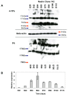
Expression analysis of pig Reelin during development. A) Western blot analysis with the monoclonal anti-Reelin antibody Clone 142 on protein samples purified from cerebral cortex of embryonic pigs at the indicated developmental stages. Brain extracts were resolved by PAGE using either a 5% (lower) or 4-15%-gradient gel (upper). Three bands are seen in the samples, of about 400, 320 and ~160 kDa in the 5% gels and 320, ~160, 70, and 65 kDa in the gradient gels. Beta-actin was used as loading control. B) Quantitative analyses of Reelin mRNA expression during pig embryonic brain development. The expression levels were normalized to the expression levels of GAPDH, 18S rRNA and β-actin in the same tissue using the geNorm program.
Figure 3
Figure 3
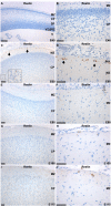
Immunohistochemical staining for Reelin in pig neocortex from five embryonic stages. A and B) Immunohistochemical staining of porcine cortex at E40. No staining was observed. C and D) Immunohistochemical staining of cortex at E60. Reelin expression is identified in CR cells of the marginal zone (MZ). B) Immunohistochemical staining at E60 at higher magnification. Black arrows indicate horizontal Reelin positive CR cells. Reelin immunoreactive interneurons in layer III-IV of the cortical plate is magnified (x10) in the box. E and F) Immunohistochemical staining at E80. Reelin expression is detectable with decreased intensity in the cortex. Bipolar fusiform CR type cells with dendrites oriented parallel with the pial surface were observed in the outer part of the MZ (open arrow) along with Reelin-immunoreactive interneurons and CR type cells displaying a variety of shapes and sizes. Reelin positive interneurons are seen in the CP but in decreased numbers. G and H) Immunoreactivity is observed in the cytoplasm of CR in the MZ at decreased numbers. The Reelin expression in the interneurons of the cortical plate is no longer detectable. I and J) Reelin positive cells are detectable in the outer part of MZ at E115. Scattered immunoreactivity is observed in the extracellular matrix throughout the CP. Cortical plate: CP; Marginal zone: MZ; pia mater: PIA; subplate: SP; VZ; subventricular zone: SVZ; 3rd ventricle: V; ventricular zone: VZ. Scale bar = 100 μm.

Similar articles

Cited by

References

    1. Angevine JB Jr, Sidman RL. Autoradiographic study of cell migration during histogenesis of cerebral cortex in the mouse. Nature. 1961;192:766–768. doi: 10.1038/192766b0. - DOI - PubMed
    1. Rakic P. Neurons in rhesus monkey visual cortex: systematic relation between time of origin and eventual disposition. Science. 1974;183(123):425–427. doi: 10.1126/science.183.4123.425. - DOI - PubMed
    1. Gleeson JG, Walsh CA. Neuronal migration disorders: from genetic diseases to developmental mechanisms. Trends Neurosci. 2000;23(8):352–359. doi: 10.1016/S0166-2236(00)01607-6. - DOI - PubMed
    1. Huang Z. Molecular regulation of neuronal migration during neocortical development. Mol Cell Neurosci. 2009;42(1):11–22. doi: 10.1016/j.mcn.2009.06.003. - DOI - PubMed
    1. D'Arcangelo G. The reeler mouse: anatomy of a mutant. Int Rev Neurobiol. 2005;71:383–417. doi: 10.1016/S0074-7742(05)71016-3. - DOI - PubMed

Publication types

MeSH terms

LinkOut - more resources