Case Reports
. 2010 Jun 15:5:36.
doi: 10.1186/1746-1596-5-36.

Invasive lobular carcinoma with extracellular mucin production and HER-2 overexpression: a case report and further case studies

Affiliations
Case Reports

Invasive lobular carcinoma with extracellular mucin production and HER-2 overexpression: a case report and further case studies

Jing Yu et al. Diagn Pathol. .

Abstract

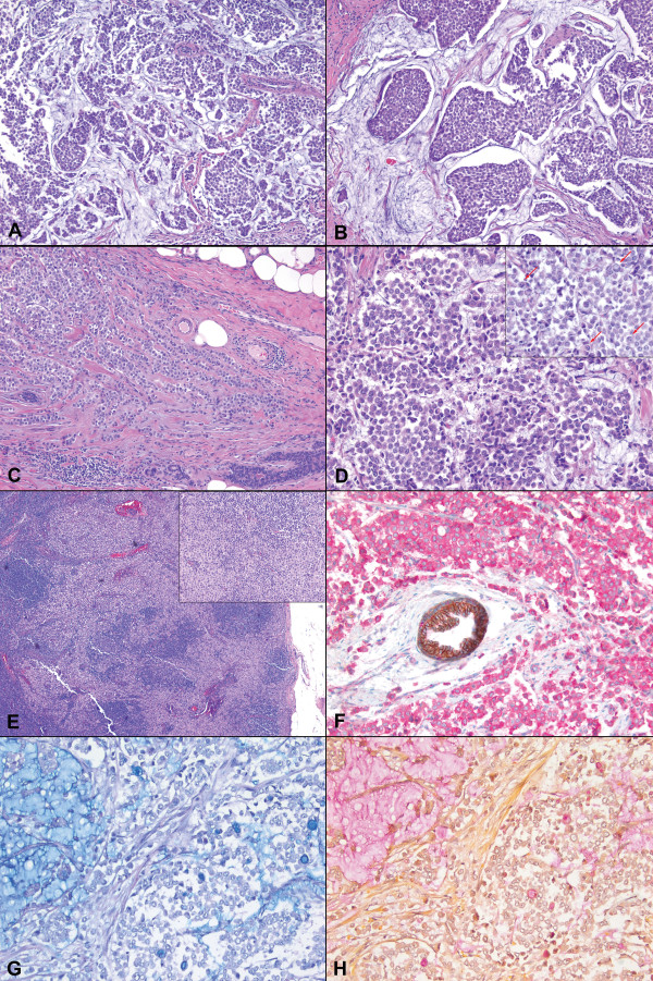
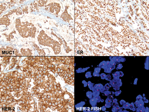
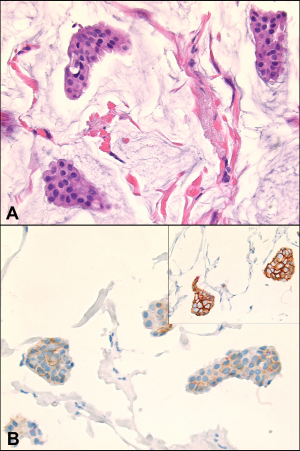
Invasive lobular carcinomas (ILC) of breast typically demonstrate intracytoplasmic mucin. We present a unique case of classical type ILC with abundant extracellular mucin and strong ERBB2 (HER2/neu) expression confirmed by immunohistochemistry and fluorescent in situ hybridization. Dual E-cadherin/p120 immunohistochemical stain demonstrated complete loss of membranous E-cadherin and the presence of diffuse cytoplasmic p120 staining, confirming the lobular phenotype. The tumor cells showed ductal-like cytoplasmic MUC1 staining, but were negative for MUC2 and other mucin gene markers. In addition, studies of tissue microarrays of 80 breast carcinomas with mucinous differentiation revealed 4 pure mucinous carcinomas showing significantly reduced E-cadherin staining without redistribution of p120 into cytoplasm. The findings suggest that the presence of extracellular mucin does not exclude a diagnosis of lobular carcinoma, and the morphologic and molecular characteristics of lobular and ductal carcinomas are more complex than previously appreciated.


Figures

Figure 1
Figure 1
A case of invasive lobular carcinoma of breast with extracellular mucin. The tumor showed discohesive (A, H&E, ×100) and nested (B, ×200) growth with abundant extracellular mucin. At the periphery, the tumor cells formed cords and single files (C, ×100). Cytologically, the tumor cells were small to medium in size and relatively uniform (D, ×400), with scattered signet ring cells present (D inset, arrows, ×400). A sentinel lymph node showed metastatic focus of uniform tumor cells (E inset, ×40) in dissociated infiltrating pattern (E, ×200). Immunohistochemical stain revealed complete absence of membranous E-cadherin staining (brown) and the presence of diffuse cytoplasmic p120 staining (pink); a normal duct with membranous staining for both E-cadherin and p120 served as an internal control (F, immunohistochemistry, ×400). Alcian Blue (G, ×200) and Mucicarmine (H, ×200) stains highlighted both extracellular and intracellular mucin.
Figure 2
Figure 2
Characteristics of the invasive lobular carcinoma with extracellular mucin. The tumor cells revealed strong cytoplasmic MUC1 staining (top left, immunohistochemistry, ×200), but were negative for MUC2, MUC4, MUC5AC and MUC6 (images not shown). The tumor cells were positive for ER (top right, ×400), negative for PR (image not shown), and positive for HER-2/neu with 3+ staining (bottom left, ×400). FISH study confirmed the amplification of HER-2 gene (bottom right, ×1,000).
Figure 3
Figure 3
Cases of pure mucinous carcinoma with significantly reduced membranous E-cadherin and p120 staining without redistribution of p120 into cytoplasm (A, H&E, ×400; B, inset is a typical mucinous carcinoma with strong membranous E-cadherin staining, immunohistochemistry, ×400).

References

    1. Gad A, Azzopardi JG. Lobular carcinoma of the breast: a special variant of mucin-secreting carcinoma. J Clin Pathol. 1975;28:711–716. doi: 10.1136/jcp.28.9.711. - DOI - PMC - PubMed
    1. Harris M, Vasudev KS, Anfield C, Wells S. Mucin-producing carcinomas of the breast: ultrastructural observations. Histopathology. 1978;2:177–188. doi: 10.1111/j.1365-2559.1978.tb01708.x. - DOI - PubMed
    1. Rosen PP. In: Rosen's Breast Pathology. 3. Rosen PP, editor. Philadelphia, PA, USA: Lippincott Williams & Wilkins; 2009. Invasive Lobular Carcinoma; pp. 690–720.
    1. Tan PH, Tse GM, Bay BH. Mucinous breast lesions: diagnostic challenges. J Clin Pathol. 2008;61:11–19. doi: 10.1136/jcp.2006.046227. - DOI - PubMed
    1. Bertucci F, Orsetti B, Negre V, Finetti P, Rouge C, Ahomadegbe JC, Bibeau F, Mathieu MC, Treilleux I, Jacquemier J, Ursule L, Martinec A, Wang Q, Benard J, Puisieux A, Birnbaum D, Theillet C. Lobular and ductal carcinomas of the breast have distinct genomic and expression profiles. Oncogene. 2008;27:5359–5372. doi: 10.1038/onc.2008.158. - DOI - PMC - PubMed

Publication types

MeSH terms