. 2010 Jun 16;98(12):2813-21.
doi: 10.1016/j.bpj.2010.03.052.

Force transmission in the organ of Corti micromachine

Affiliations

Force transmission in the organ of Corti micromachine

Jong-Hoon Nam et al. Biophys J. .

Abstract

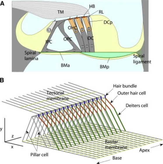
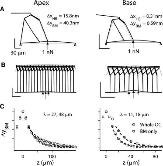
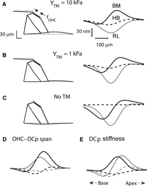
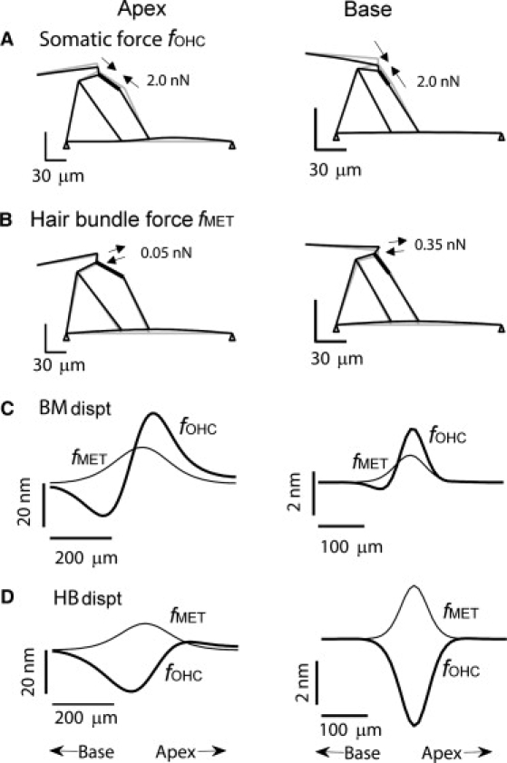
Auditory discrimination is limited by the performance of the cochlea whose acute sensitivity and frequency tuning are underpinned by electromechanical feedback from the outer hair cells. Two processes may underlie this feedback: voltage-driven contractility of the outer hair cell body and active motion of the hair bundle. Either process must exert its mechanical effect via deformation of the organ of Corti, a complex assembly of sensory and supporting cells riding on the basilar membrane. Using finite element analysis, we present a three-dimensional model to illustrate deformation of the organ of Corti by the two active processes. The model used available measurements of the properties of structural components in low-frequency and high-frequency regions of the rodent cochlea. The simulations agreed well with measurements of the cochlear partition stiffness, the longitudinal space constant for point deflection, and the deformation of the organ of Corti for current injection, as well as displaying a 20-fold increase in passive resonant frequency from apex to base. The radial stiffness of the tectorial membrane attachment was found to be a crucial element in the mechanical feedback. Despite a substantial difference in the maximum force generated by hair bundle and somatic motility, the two mechanisms induced comparable amplitudes of motion of the basilar membrane but differed in the polarity of their feedback on hair bundle position. Compared to the hair bundle motor, the somatic motor was more effective in deforming the organ of Corti than in displacing the basilar membrane.


Figures

Figure 1
Figure 1
Finite element model of cochlear partition. (A) Radial section of cochlear partition. Organ of Corti sits on basilar membrane (BM), which is divided into arcuate (BMA) and pectinate (BMP) zones. BMA supports inner and outer pillar cells (IPC and OPC) and BMP supports Deiters' cell (DC) and outer hair cell (OHC). Reticular lamina (RL) comprises tops of pillar cells, OHC cuticular plates, and tops of DC phalangeal processes (DCP). (B) Structural components of cochlear partition represented by beam elements allowing elongation and bending. The medial and lateral ends of the basilar membrane were hinged at the spiral lamina and spiral ligament and the tectorial membrane (TM) was fixed. Both elements were composed of beam elements aligned in radial and longitudinal directions. Three rows of OHCs and DCs merged into one with Young's modulus and force generation three-times that of single cell. Deiters' cell phalangeal process (DCP) and OHC form two arms of a Y-shape, surmounting the basal part of the DC. Scale bars 40 μm.
Figure 2
Figure 2
Deformation due to point load at cochlear apex and base. (A) Radial section of organ of Corti showing deformation by 1 nN of force applied to basilar membrane beneath Deiters'cell. Scale bars 30 μm. Deformation exaggerated for illustration. (B) Longitudinal section showing force is distributed over 30 μm, OHCs 10 μm separation. Note Y-shaped structure of OHC (thick line) and Deiters cell. (C) Plots of basilar membrane displacement, ΔyBM, normalized to maximum value, ΔyBM-MAX, along the longitudinal direction for entire cochlear partition (circles) and basilar membrane alone without the organ of Corti (squares). Stiffness of the partition and the longitudinal space constant, λ, for the apex were 0.025 N/m and 48 μm, which were reduced to 0.005 N/m and 27 μm when only basilar membrane was simulated. Stiffness and λ for base were 1.69 N/m and 18 μm, which were reduced to 0.77 N/m and 11 μm with basilar membrane alone.
Figure 3
Figure 3
Deformation of the organ of Corti by OHC somatic motor depends on mechanical properties. Effects of variations in tectorial membrane stiffness (AC) and tilt and stiffness of Deiters' cell phalangeal process (DCP) (D and E). OHCs stimulated to produce an axial contractile force (fOHC) of 2 nN per OHC distributed longitudinally with standard deviation σz = 60 μm. The cochlear apex was simulated for three different values of tectorial membrane body radial stiffness with Young's modulus, YTM, of 10 kPa (A), 1 kPa (B), and 0 kPa (C). (Left) Radial sections; (right) longitudinal profile of the displacements of the basilar membrane (BM; thick line), hair bundle (HB, dashed line), and reticular lamina (RL, thin line), each at a radial position beneath the base of the DC. Scale bars 30 μm. The radial profile in panel B resembles that measured in Karavitaki and Mountain (37). (D) Effects on longitudinal profile of displacement of the basilar membrane (thick line) and hair bundle (dashed line) for changes in OHC-DC longitudinal span, 40 μm (four OHC diameters; black), and 0 μm (no separation; gray) where default in panel A is 20 μm (two OHC diameters). (E) Change in DC-phalangeal process stiffness, 0.05 MPa (black), 5 MPa (gray) where default is 5 MPa. In panels AC, the OHC-DC span was 20 μm with OHCs tilted 5 μm toward the base. In panels D and E, Young's modulus of the tectorial membrane body was 10 kPa.
Figure 4
Figure 4
Static displacements induced by OHC active forces. Radial sections of the organ of Corti showing motion produced by (A) an OHC somatic (contractile) force (fOHC) or (B) a hair bundle force (fMET). Forces distributed longitudinally with σz = 60 μm (apex) and 25 μm (base) with peak values of fOHC = 2.0 nN (base and apex) and fMET = 0.05 nN (apex) or 0.35 nN (base). Longitudinal profiles of the basilar membrane (BM) displacement (C) and hair bundle (HB) displacement (D) for application of the active forces fOHC (thick lines) and fMET (thin lines). Displacement determined at a radial position beneath the base of the DC. (Left column) Apex. (Right column) Base.
Figure 5
Figure 5
Dynamic responses of the organ of Corti to current injection. OHCs stimulated to produce an axial contractile force (fOHC) of 1 nN peak value distributed longitudinally with standard deviation σz = 60 μm at the base (A) and apex (B and C). (A) Base: time course of dynamic displacements of the basilar membrane (BM, thick line), hair bundle (HB, dashed line), and reticular lamina (RL, thin line) for a force-step timing that is shown in the top trace. (B) Apex: time courses of displacements for the same force step. (C) Apex: effects of a 1000-fold reduction in tectorial membrane attachment stiffness. In panel C, the hair bundle shows damped oscillations at a frequency (1.4 kHz) higher than the basilar membrane, determined by tectorial membrane mass and hair bundle stiffness. In the standard conditions, the basilar membrane displays damped oscillations at 0.7 kHz (apex) and 16 kHz (base).

Similar articles

Cited by

References

    1. Angelborg C., Engström H. Supporting elements in the organ of Corti. I. Fibrillar structures in the supporting cells of the organ of Corti of mammals. Acta Otolaryngol. Suppl. 1972;301:49–60. - PubMed
    1. Voldřich L. Experimental and topographic morphology in cochlear mechanics. In: de Boer E., Viergever M.A., editors. Mechanics of Hearing. Delft University Press; Delft, The Netherlands: 1983.
    1. Slepecky N.B. Structure of the mammalian cochlea. In: Dallos P., Popper A.N., Fay R.R., editors. The Cochlea. Springer; New York: 1996.
    1. Ashmore J. Cochlear outer hair cell motility. Physiol. Rev. 2008;88:173–210. - PubMed
    1. Hudspeth A. Mechanical amplification of stimuli by hair cells. Curr. Opin. Neurobiol. 1997;7:480–486. - PubMed

Publication types

LinkOut - more resources