. 2010 Jun 16;98(12):2923-32.
doi: 10.1016/j.bpj.2010.03.023.

Mechanics of Vorticella contraction

Affiliations

Mechanics of Vorticella contraction

Gaurav Misra et al. Biophys J. .

Abstract

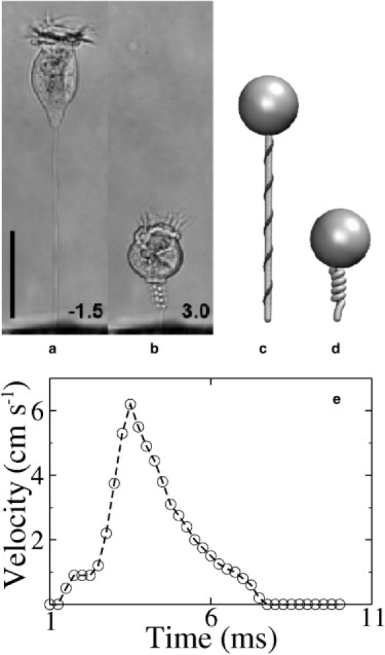
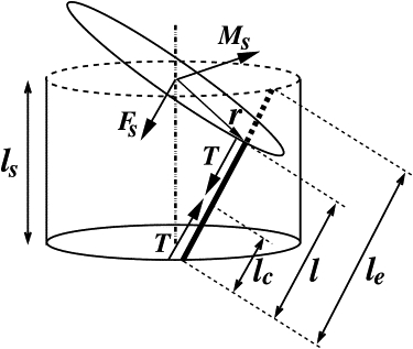
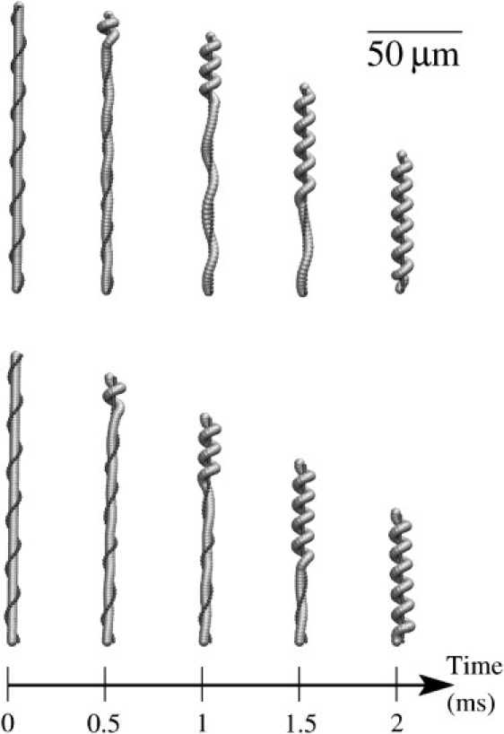
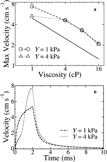
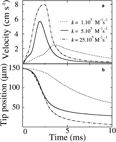
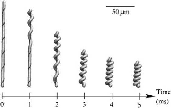
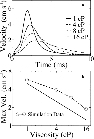
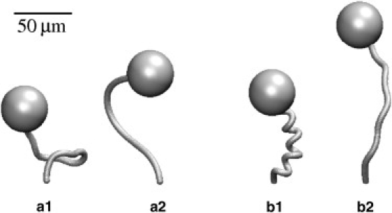
Vorticella convallaria is one of a class of fast-moving organisms that can traverse its body size in less than a millisecond by rapidly coiling a slender stalk anchoring it to a nearby surface. The stalk houses a fiber called the spasmoneme, which winds helically within the stalk and rapidly contracts in response to calcium signaling. We have developed a coupled mechanical-chemical model of the coiling process, accounting for the coiling of the elastic stalk and the binding of calcium to the protein spasmin. Simulations of the model describe the contraction and recovery processes quantitatively. The stalk-spasmoneme system is shown to satisfy geometric constraints, which explains why the cell body sometimes rotates during contraction. The shape of the collapsing and recovering stalk bounds its effective bending stiffness. Simulations suggest that recovery from the contracted state is driven by the stalk at a rate controlled by dissociation of calcium from spasmin.


Figures

Figure 1
Figure 1
(a and b) Images of V. convallaria in extended and contracted states (22), reproduced with permission. (c and d) The model in extended and contracted states. The head is modeled by an incompressible sphere, the stalk by an elastic rod (in gray), and the spasmoneme by a thin fiber (in black) winding helically around the stalk. (e) A typical velocity profile of the cell body is shown from the time when the motion starts; the graph was redrawn from Fig. 2 of Upadhyaya et al. (25). The scale bar in panels ad is 50 μm.
Figure 2
Figure 2
A segment of the rod-fiber assembly showing the tension (T) in the fiber and its position relative to the centerline of the rod. The segment length in the discretized model is given by ls = L/Ns where L is the length of the rod and Ns is the number of segments. The force acting on the centerline (Fs) is parallel to T and exerts shear and compression forces. The moment (Ms) of the tension is perpendicular to r and Fs, and generates bend and twist couples. The rest length of the spasmoneme segment in its calcium-free state is le and its rest length in the calcium-bound state is lc; its time-dependent length, l, varies between le and lc. The force and couple acting on the bottom plane are not shown.
Figure 3
Figure 3
Time-lapse images of the initial phase of contraction driven by a propagating calcium signal (for clarity, the head is not shown). (Top) Young's modulus Y = 1 kPa. (Bottom) Y = 4 kPa.
Figure 4
Figure 4
Simulation results for contraction driven by a propagating calcium signal. (a) Peak velocity versus viscosity. The solid line indicates a slope of –0.5. (b) Velocity profiles. The peak velocity decays more rapidly than the experimental data shown in Fig. 1e. For the stiffer stalk (Y = 4 kPa), the velocity increases much too rapidly as well.
Figure 5
Figure 5
Results for Young's modulus Y = 1 kPa and different values of the rate constant, k. (a) Velocity of the head versus time. (b) Position of the head versus time.
Figure 6
Figure 6
Time-lapse images of contraction with Young's modulus Y = 1 kPa and rate constant k = 5 × 10⁵ M−1 s−1.
Figure 7
Figure 7
Velocity data for Young's modulus Y = 1 kPa and rate constant k = 5 × 10⁵ M−1 s−1. (a) Velocity profile for different viscosities. (b) Peak velocity versus viscosity. The solid line depicts the slope of –0.5.
Figure 8
Figure 8
Recovery simulations with Young's modulus Y = 0.25 kPa (a1 and a2) and Y = 1 kPa (b1 and b2). Early stalk shapes are shown in a1 and b1, while a2 and b2 show the shapes at a later time.

Similar articles

Cited by

References

    1. Mahadevan L., Matsudaira P. Motility powered by supramolecular springs and ratchets. Science. 2000;288:95–99. - PubMed
    1. Amos W.B. Structure and coiling of the stalk in the peritrich ciliates Vorticella and Carchesium. J. Cell Sci. 1972;10:95–122. - PubMed
    1. Allen R.D. Structures linking the myonemes, endoplasmic reticulum, and surface membranes in the contractile ciliate Vorticella. J. Cell Biol. 1973;56:559–579. - PMC - PubMed
    1. Hoffmannberling H. The mechanism of a novel contraction cycle that differs from the muscle contraction. [Der mechanismus eines neuen, von der muskelkontraktion verschiedenen kontraktionszyklus] Biochim. Biophys. Acta. 1958;27:247–255. - PubMed
    1. Amos W.B. Reversible mechanochemical cycle in the contraction of Vorticella. Nature. 1971;229:127–128. - PubMed

Publication types

LinkOut - more resources