. 2010 Jun 16;98(12):3044-53.
doi: 10.1016/j.bpj.2010.03.040.

Hydrodynamic properties of human adhesion/growth-regulatory galectins studied by fluorescence correlation spectroscopy

Affiliations

Hydrodynamic properties of human adhesion/growth-regulatory galectins studied by fluorescence correlation spectroscopy

Antonia Göhler et al. Biophys J. .

Abstract

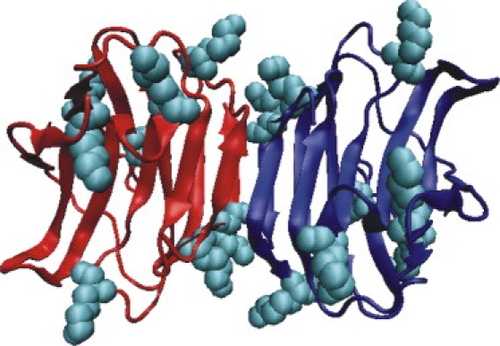
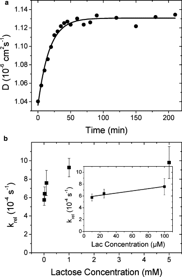
Fluorescence correlation spectroscopy is applied on homologous human lectins (i.e., adhesion/growth-regulatory galectins) to detect influence of ligand binding and presence of the linker peptide in tandem-repeat-type proteins on hydrodynamic properties. Among five tested proteins, lactose binding increased the diffusion constant only in the cases of homodimeric galectin-1 and the linkerless variant of tandem-repeat-type galectin-4. To our knowledge, the close structural similarity among galectins does not translate into identical response to ligand binding. Kinetic measurements show association and dissociation rate constants in the order of 1 to 10(3) M(-1) s(-1) and 10(-4) s(-1), respectively. Presence of the linker peptide in tandem-repeat-type protein leads to anomalous scaling with molecular mass. These results provide what we believe to be new insights into lectin responses to glycan binding, detectable so far only by small angle neutron scattering, and the structural relevance of the linker peptide. Methodologically, fluorescence correlation spectroscopy is shown to be a rather simple technical tool to characterize hydrodynamic properties of these proteins at a high level of sensitivity.


Figures

Figure 1
Figure 1
Illustration of the three types of spatial arrangement of carbohydrate recognition domains in human galectins using the tested representatives as examples: homodimeric prototype galectin-1 (top; hGal-1), chimera-type galectin-3 with its N-terminal domain for serine phosphorylation and its collagenase-sensitive stalk (middle; hGal-3) and tandem-repeat-type galectins-4, -8, and -9 (bottom; hGal-4, hGal-8/-9). Two tested variants include proteolytically truncated galectin-3 (hGal-3tr) and the conversion of tandem-repeat-type galectin-4 into a prototype form by loss of its linker peptide (hGal-4PT). Ligand binding is given, its effect on the diffusion constant is reflected in the size of the depicted lectin (compaction) and the color toning.
Figure 2
Figure 2
Crystal structure of homodimeric hGal-1 (PDB code: 1gzw) displayed using VMD (visualization of protein structure with protomer 1 in red, protomer 2 in blue). All lysine residues are given as van der Waals spheres (cyan).
Figure 3
Figure 3
Fluorescence labeling of hGal-1. (a) Absorption spectra of hGal-1 labeled with ALEXA647 with a DOL≈1 (black) and a DOL≈1.3 (gray) in comparison to that of the freely diffusing, hydrolyzed fluorophore ALEXA647 (light gray). The increase in extent of extinction at ∼610 nm (arrow) at increased DOL due to fluorophore dimerization is clearly visible. (b) Exemplary FCS data (black line) for ALEXA647-labeled hGal-1. The decay on the μs-timescale (left arrow) is due to fluorophore photophysics, whereas the ms-decay (right arrow) represents translational diffusion of the protein. The data is fitted according to Eq. 2 (gray line).
Figure 4
Figure 4
Diffusion constant D for hGal-1 measured by FCS at various concentrations of lactose. The binding curve was fitted by a Hill function (Eq. 6) yielding results as listed in Table 1. (Top) Average brightness per molecule B for hGal-1 estimated from each corresponding FCS measurement. The straight line represents the average value of B over all measurements. All error bars represent SE of 0.5%.
Figure 5
Figure 5
Diffusion constant D for (a) hGal-4PT, the engineered hGal-4 variant without linker, and (b) hGal-4 measured by FCS at various concentrations of lactose. The binding curve in the left graph was fitted by a Hill function (Eq. 6) yielding results as listed in Table 1. (Top) The corresponding brightness per molecule B. Straight lines represent the average value of B over all measurements. All error bars represent SE estimated from repeated measurements.
Figure 6
Figure 6
Kinetics of binding of lactose to hGal-1. (a) Diffusion constants D (circles) of hGal-1 in the presence of 5 mM lactose as function of time and fitted with an exponential rise function yielding a relaxation rate constant of (9.6 ± 0.7) × 10−4 s−1 (line). (b) Experimentally derived rate constants krel as function of lactose concentration clac (squares). Error bars represent uncertainties as estimated from the nonlinear fit routine in Origin software. (Inset) Experimentally derived rate constant krel for lactose concentrations clac < 100 μM fitted with a linear function (line) yielding an association rate constant (slope) of (2.0 ± 0.6) s−1 M−1 and a dissociation rate constant (offset) of (5.7 ± 0.2) × 10−4 s−1.

References

    1. Gabius H.-J., editor. The Sugar Code. Fundamentals of Glycosciences. Wiley-VCH; Weinheim: 2009.
    1. Villalobo A., Nogales-González A., Gabius H.-J. A guide to signaling pathways connecting protein-glycan interaction with the emerging versatile effector functionality of mammalian lectins. Trends Glycosci. Glycotechnol. 2006;18:1–37.
    1. Gabius H.-J. Glycans: bioactive signals decoded by lectins. Biochem. Soc. Trans. 2008;36:1491–1496. - PubMed
    1. Wu A.M., Wu J.H., Gabius H.-J. Effects of polyvalency of glycotopes and natural modifications of human blood group ABH/Lewis sugars at the Galβ1-terminated core saccharides on the binding of domain-I of recombinant tandem-repeat-type galectin-4 from rat gastrointestinal tract (G4-N) Biochimie. 2004;86:317–326. - PubMed
    1. André S., Kožár T., Gabius H.-J. Substitutions in the N-glycan core as regulators of biorecognition: the case of core-fucose and bisecting GlcNAc moieties. Biochemistry. 2007;46:6984–6995. - PubMed

Publication types