. 2010 Jun 16;98(12):3070-7.
doi: 10.1016/j.bpj.2010.02.047.

Two-dimensional nanoscale structural and functional imaging in individual collagen type I fibrils

Affiliations

Two-dimensional nanoscale structural and functional imaging in individual collagen type I fibrils

Catalin Harnagea et al. Biophys J. .

Abstract

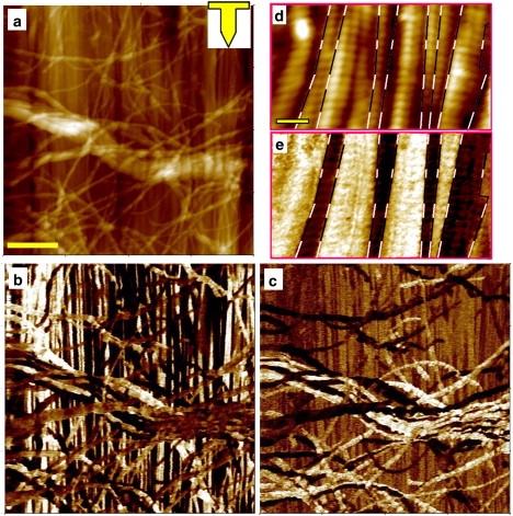
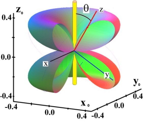
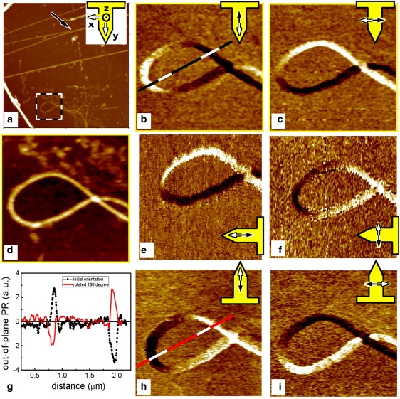
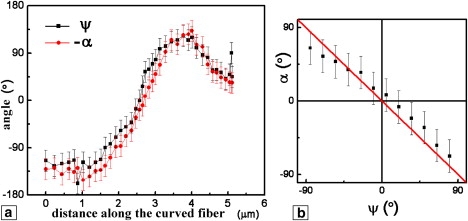
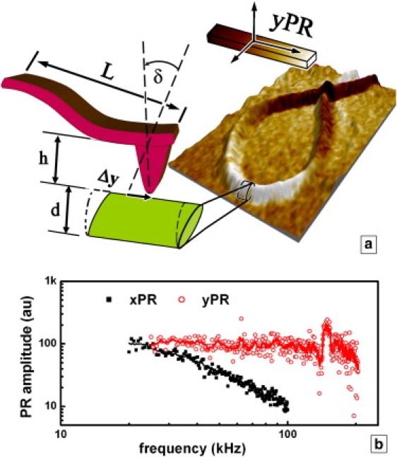
The piezoelectric properties of single collagen type I fibrils in fascia were imaged with sub-20 nm spatial resolution using piezoresponse force microscopy. A detailed analysis of the piezoresponse force microscopy signal in controlled tip-fibril geometry revealed shear piezoelectricity parallel to the fibril axis. The direction of the displacement is preserved along the whole fiber length and is independent of the fiber conformation. It is shown that individual fibrils within bundles in skeletal muscle fascia can have opposite polar orientations and are organized into domains, i.e., groups of several fibers having the same polar orientation. We were also able to detect piezoelectric activity of collagen fibrils in the high-frequency range up to 200 kHz, suggesting that the mechanical response time of biomolecules to electrical stimuli can be approximately 5 micros.


Figures

Figure 1
Figure 1
AFM topography images (a and d) and the corresponding PR images of the fascia: VPR (b and e) and LPR (c). The inset in a shows the cantilever orientation during the measurement. The scale bar in a is 2 μm (valid for images a–c). Higher-magnification images (d and e) clearly show the 67 nm periodicity and the organization of fibrils into domains (the domain boundaries are marked with dashed lines). The scale bar in d is 250 nm (valid for images d and e).
Figure 2
Figure 2
The longitudinal piezoelectric coefficient dzz along different directions (the region [x0 > 0, y0 < 0] is removed). The coefficient nullifies for θ = 90° (dzz = d11 = d22 = 0).
Figure 3
Figure 3
PR of an isolated collagen fiber at three cantilever orientations with respect to the sample: at an initial, arbitrary orientation (b and c); after a 90° clockwise rotation (e and f); and at 180° orientation (h and i). Images b, e, and h are constructed from the out-of-plane deflection signal, and c, f, and i represent the LPR. The topography of the area is shown in a (10 μm scan) and d (2 μm zoom of a). The plot in g shows the change in VPR along the same line of the sample (b and h) before and after the sample has been rotated by 180°. The cantilever orientation and the detection direction are overlaid on the PFM images.
Figure 4
Figure 4
Comparison of the orientation of the fiber and the in-plane PR vector with respect to the cantilever axis. (a) Angles calculated from Fig. 2, b–d. (b) Independent measurement obtained on a long, straight fiber by rotating the sample in increments of 15°; calculated angle between the PR vector and the cantilever axis versus the angle between the straight fiber and the cantilever.
Figure 5
Figure 5
(a) Schematic representation of the VPR detection in PFM using the cantilever buckling. The fiber in Fig 2 is shown in a three-dimensional view, with the color scale being its yPR (Fig. 2b). Main physical quantities involved: L, cantilever length; h, tip height; δ, cantilever deflection angle; Δy, sample in-plane displacement; and d, sample thickness. (b) Frequency dependence of the amplitude of the VPR and LPR signals normalized to the same low-frequency value.

References

    1. Ramachandra G.N. Structure of Collagen at the Molecular Level. In: Ramachandran G.N., editor. Treatise on Collagen: Chemistry of Collagen. Vol. 1. Academic Press; New York: 1967.
    1. Kadler K.E., Hill A., Canty-Laird E.G. Collagen fibrillogenesis: fibronectin, integrins, and minor collagens as organizers and nucleators. Curr. Opin. Cell Biol. 2008;20:495–501. - PMC - PubMed
    1. Kühn K. The structure of collagen. Essays Biochem. 1969;5:59–87. - PubMed
    1. Baselt D.R., Revel J.P., Baldeschwieler J.D. Subfibrillar structure of type I collagen observed by atomic force microscopy. Biophys. J. 1993;65:2644–2655. - PMC - PubMed
    1. Paige M.F., Rainey J.K., Goh M.C. Fibrous long spacing collagen ultrastructure elucidated by atomic force microscopy. Biophys. J. 1998;74:3211–3216. - PMC - PubMed

Publication types

Substances

LinkOut - more resources