. 2010 Jun 11;141(6):970-81.
doi: 10.1016/j.cell.2010.04.038.

ATM-dependent chromatin changes silence transcription in cis to DNA double-strand breaks

Affiliations

ATM-dependent chromatin changes silence transcription in cis to DNA double-strand breaks

Niraj M Shanbhag et al. Cell. .

Abstract

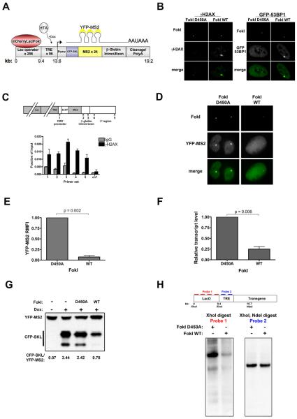
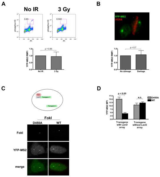
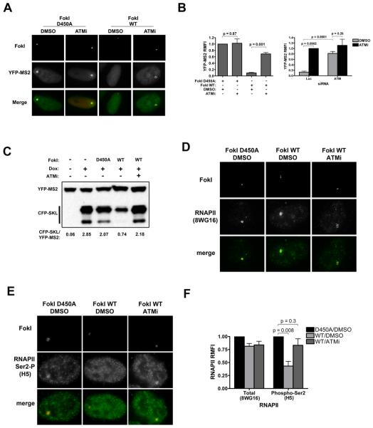
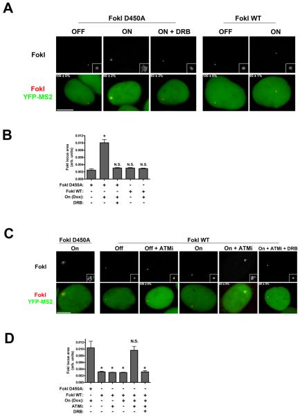
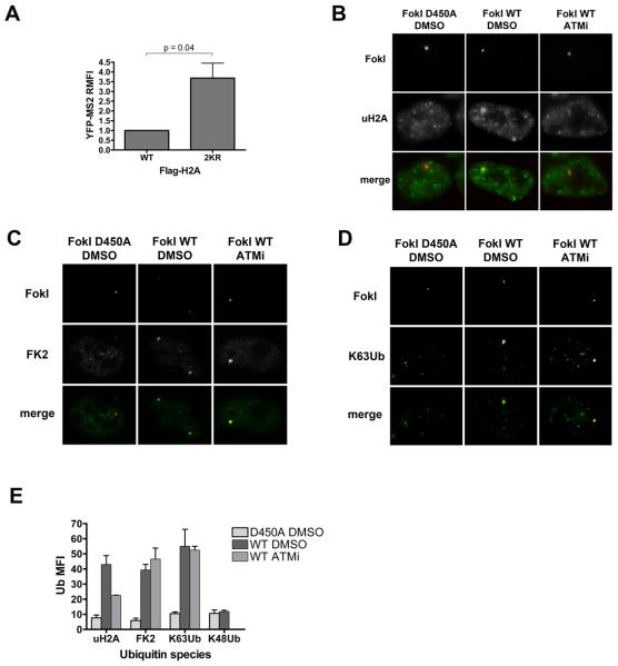
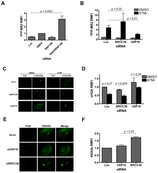
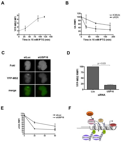
DNA double-strand breaks (DSBs) initiate extensive local and global alterations in chromatin structure, many of which depend on the ATM kinase. Histone H2A ubiquitylation (uH2A) on chromatin surrounding DSBs is one example, thought to be important for recruitment of repair proteins. uH2A is also implicated in transcriptional repression; an intriguing yet untested hypothesis is that this function is conserved in the context of DSBs. Using a novel reporter that allows for visualization of repair protein recruitment and local transcription in single cells, we describe an ATM-dependent transcriptional silencing program in cis to DSBs. ATM prevents RNA polymerase II elongation-dependent chromatin decondensation at regions distal to DSBs. Silencing is partially dependent on E3 ubiquitin ligases RNF8 and RNF168, whereas reversal of silencing relies on the uH2A deubiquitylating enzyme USP16. These findings give insight into the role of posttranslational modifications in mediating crosstalk between diverse processes occurring on chromatin.


Figures

Figure 1
Figure 1. DSBs initiate local transcriptional silencing
(A) Schematic of reporter locus. 256 lac operator repeats and a tetracycline response element (TRE) array are upstream of an inducible reporter gene that codes for the CFP-SKL protein and contains 24 repeats of the MS2 RNA stem loop sequence. The lac repressor is fused to mCherry and to the nuclease domain of the FokI endonuclease, enabling DSB induction within the 9 kb of lac operator repeats. Nascent transcript is visualized through binding of a YFP-MS2 protein to the MS2 stem loops. CFP-SKL accumulates in the cytoplasm upon induction of transcription. Not drawn to scale. (B) Transfection of the wild type FokI nuclease fusion (FokI WT) but not the nuclease-deficient D450A mutant into U2OS cells containing the stably integrated reporter leads to local accumulation of damage response proteins γH2AX (left) and 53BP1 (right). (C) Top – schematic of reporter locus and primers used for chromatin immunoprecipitation (ChIP). Bottom – ChIP analysis demonstrates enrichment of γH2AX across reporter locus compared to IgG control. ch7 represents negative control locus on chromosome 7. Data are means + standard error of the mean (SEM) from two independent experiments. (D) Nascent transcript (YFP-MS2) accumulation in cells expressing FokI D450A and FokI WT. (E) Quantification of YFP-MS2 relative mean fluorescence intensity (RMFI) from (D) using ImageJ software (NIH). Data was derived from 3 independent experiments and is depicted relative to FokI D450A expressing cells. Error bars represent SEM. (F) qRT-PCR for reporter mRNA levels in cells 48 hours after transduction with lentivirus expressing FokI WT or D450A. Transcription was monitored 3 hours after addition of 1 μg/ml doxycycline. Error bars represent SEM from 3 independent experiments. (G) Representative immunoblot (IB) for CFP-SKL in cells expressing FokI WT or D450A after 4 hr induction with 1 μg/ml doxycycline. The ratio of CFP-SKL to YFP-MS2 signal density was quantified using ImageJ software (NIH). (H) Top – schematic of Southern blot strategy. Bottom - Southern blot of genomic DNA from reporter cells expressing FokI WT or D450A digested with restriction enzymes XhoI, or XhoI and NdeI and probed for the indicated regions. See also Figure S1.
Figure 2
Figure 2. DSB induced silencing occurs in cis to DNA damage
(A) Top – flow cytometric analysis of reporter cells mock treated (No IR) or treated with 3 Gy of ionizing radiation (IR), and analyzed for phospho-histone H3 and propidium iodide after 3 hours. Mitotic cells are contained within the red box and the percentage of mitotic cells indicated. Bottom – quantification of YFP-MS2 RMFI in mock and 3 Gy treated cells after 3 hour induction with 1 μg/ml doxycycline. The experiment was performed in triplicate and data represent mean + standard deviation (SD). (B) Cells were induced with doxycycline for 3 hrs immediately after laser microirradiation. Top – representative image of microirradiated cell (right) identified by γH2AX (red) and a neighboring, undamaged cell. Nascent transcript is visible as YFP-MS2 in both cells. Bottom – Quantification of YFP-MS2 fluorescence intensity in damaged and undamaged cells. The experiment was performed in triplicate and data represent mean + SD. (C) Top – schematic of two transgene containing reporter cells. Both transgenes contain identical promoters and regulatory elements, while only one contains the lac operator array. Bottom – representative images of two transgene reporter cells expressing FokI D450A (left) or WT (right), and YFP-MS2 accumulation at both transgenes. (D) Quantification of YFP-MS2 intensity at both arrays as in (C). Data are presented as mean + SEM from two independent experiments. See also Figure S2.
Figure 3
Figure 3. Transcriptional silencing at DSBs is dependent on ATM kinase activity
(A) Reporter cells expressing FokI D450A or FokI WT were treated with 1μg/ml dox and either DMSO or ATMi (10 μM KU55933 ) for 3 hours before fixing for analysis of YFP-MS2 accumulation. (B) Left - Quantification of YFP-MS2 RMFI from experiments as in (A). Error bars represent SEM from two to four independent experiments. Right – quantification of RMFI in cells expressing FokI WT, treated with siRNA against control or ATM, and treated with DMSO or ATMi. Error bars represent SEM from 2 independent experiments. (C) Representative IB for CFP-SKL protein in D450A or FokI WT expressing cells treated with either DMSO or ATMi and 1μg dox for 4 hours. YFP-MS2 levels are used as a loading control. (D) IF for total RNAPII (8WG16) was performed as indicated in cells treated with DMSO or with ATMi. (E) IF was performed for each group as in (D) for actively elongating RNAPII with an antibody to RNAPII phosphorylated on Ser2 of the C-terminal domain (H5). (F) Quantification of RMFI from experiments in panels D and E. Error bars represent SEM from at least three independent experiments. See also Figure S3.
Figure 4
Figure 4. ATM prevents transcription-dependent large-scale chromatin decondensation at sites of DSBs
(A) Representative images of mCherry-FokI loci in various transcriptional states with or without DSB induction. Numbers in bottom panels represent percentage of cells with the YFP-MS2 accumulation seen in the image + SEM. Insets in top panels are equivalent magnifications. Bar, 10 μM. (B) Quantification of mCherry-FokI locus size from experiments as in (A). Representative cells were imaged for quantification based on YFP-MS2 signal. Bars represent mean + SEM from 3 independent experiments. (C) Representative images of mCherry-FokI loci in various transcriptional states with or without DSB induction. Numbers in bottom panels represent percentage of cells with the YFP-MS2 accumulation seen in the image ± SEM. Insets in top panels are equivalent magnifications. Bar, 10 μM. (D) Quantification of mCherry-FokI locus size from experiments as in (B). Representative cells were imaged for quantification based on YFP-MS2 signal. Bars represent mean + SEM from 3 independent experiments. See also Figure S4.
Figure 5
Figure 5. ATM-dependent transcriptional silencing is associated with H2A ubiquitylation
(A) YFP-MS2 RMFI was quantified in cells expressing FokI WT and either Flag-H2A (WT) or a Flag-H2A allele mutated at lysines 119 and 120 to arginines (2KR). Error bars represent SEM from 4 independent experiments. (B-D) Representative images of uH2A (B), conjugated polyubiquitin (FK2) (C), and K63Ub (D) in the presence of FokI D450A or FokI WT and DMSO or ATMi. (E) Quantification of Ub MFI from experiments as in B-D. Error bars indicate SEM from three independent experiments. See also Figure S5.
Figure 6
Figure 6. Opposing regulation of DSB induced transcriptional silencing by E3 ligases RNF8 and RNF168, and the deubiquitylating enzyme USP16
(A) YFP-MS2 intensity analysis was performed 48-72 hours after transduction of cells with lentivirus expressing FokI WT and transfection with the indicated siRNAs. Error bars represent SEM from at least 3 independent experiments. (B) YFP-MS2 accumulation was assessed in cells transduced with lentivirus expressing FokI WT and treated with siRNA against luciferase (Luc) or the deubiquitylating enzymes USP16 and BRCC36 (B36). Transcription was induced with 1 μg/ml doxycycline and either vehicle or 10 μM ATMi for 3 hours. Error bars represent SEM from at least 3 independent experiments. (C) Representative images of uH2A (green) at FokI-induced DSBs (red) in cells transfected with siRNAs targeted to luciferase (Luc), USP16, or BRCC36 (B36) 48-72 hours prior to treatment with vehicle or 10 μM ATMi for 3 hours. (D) Quantification of RMFI from experiments as in C. Error bars represent SEM from 3 independent experiments. (E) Representative images of K63Ub at FokI-induced DSBs in cells transfected with the indicated siRNAs 48-72 hours prior to a 3 hour treatment with vehicle or 10 μM ATMi. (F) Quantification of RMFI from experiments as in E. Error bars represent SEM from 2 independent experiments. See also Figure S6.
Figure 7
Figure 7. DSB repair leads to rapid transcriptional derepression that is dependent on USP16
(A) Reporter cells expressing FokI WT were treated with 15 mM IPTG and a time course of nascent transcription was assessed by accumulation of YFP-MS2. Treatments were all timed to end after 3 hours of treatment with 1 μg/ml doxycycline. Error bars represent SD from duplicate experiments. (B) Loss of DSB associated ubiquitin species was monitored following IPTG treatment by performing IF with antibodies to uH2A or K63Ub at breaks. Because IPTG was used, a mCherry transactivator was used to locate the locus. Error bars represent SD from duplicate experiments. (C) Reporter cells were transfected with siRNA to luciferase (Luc) or USP16 and transduced with FokI expressing lentivirus 24 hours later. 48 hours after lentivirus transduction, cells were treated with 1 μg doxycycline and 15 mM IPTG for 3 hours. Representative images are shown. FokI images (top) are enhanced to demonstrate pan-nuclear localization upon IPTG treatment. (D) Quantification of YFP-MS2 accumulation from experiments as in D. Error bars represent SEM from 2 independent experiments. (E) Loss of uH2A at reporter locus over time in 15mM IPTG in cells expressing FokI WT and treated with control or siRNA against USP16. Because IPTG was used, a mCherry transactivator was used to locate the locus. Error bars represent SEM from 3 independent experiments. (F) ATM dependent localization of RNF8 and RNF168 to DSBs facilitates K63-ubiquitylation of one or more as yet undefined substrates (X) as well as H2A ubiquitylation (gray cylinders with a single oval ubiquitin). K63Ub attracts the BRCA1-RAP80 complex, while uH2A mediates transcriptional silencing on chromatin spanning multiple kb in cis to the DSB. USP16 deubiquitylates uH2A to restore transcription following ATMi or cessation of damage.

Comment in

References

    1. Anindya R, Aygun O, Svejstrup JQ. Damage-induced ubiquitylation of human RNA polymerase II by the ubiquitin ligase Nedd4, but not Cockayne syndrome proteins or BRCA1. Mol Cell. 2007;28:386–397. - PubMed
    1. Bartkova J, Horejsi Z, Koed K, Kramer A, Tort F, Zieger K, Guldberg P, Sehested M, Nesland JM, Lukas C, et al. DNA damage response as a candidate anti-cancer barrier in early human tumorigenesis. Nature. 2005;434:864–870. - PubMed
    1. Bredemeyer AL, Helmink BA, Innes CL, Calderon B, McGinnis LM, Mahowald GK, Gapud EJ, Walker LM, Collins JB, Weaver BK, et al. DNA double-strand breaks activate a multi-functional genetic program in developing lymphocytes. Nature. 2008;456:819–823. - PMC - PubMed
    1. Bregman DB, Halaban R, van Gool AJ, Henning KA, Friedberg EC, Warren SL. UV-induced ubiquitination of RNA polymerase II: a novel modification deficient in Cockayne syndrome cells. Proc Natl Acad Sci U S A. 1996;93:11586–11590. - PMC - PubMed
    1. Cooper EM, Cutcliffe C, Kristiansen TZ, Pandey A, Pickart CM, Cohen RE. K63-specific deubiquitination by two JAMM/MPN+ complexes: BRISC-associated Brcc36 and proteasomal Poh1. Embo J. 2009;28:621–631. - PMC - PubMed

Publication types