. 2010 Jun 11;141(6):1006-17.
doi: 10.1016/j.cell.2010.04.040.

A mammalian siderophore synthesized by an enzyme with a bacterial homolog involved in enterobactin production

Affiliations

A mammalian siderophore synthesized by an enzyme with a bacterial homolog involved in enterobactin production

Laxminarayana R Devireddy et al. Cell. .

Abstract

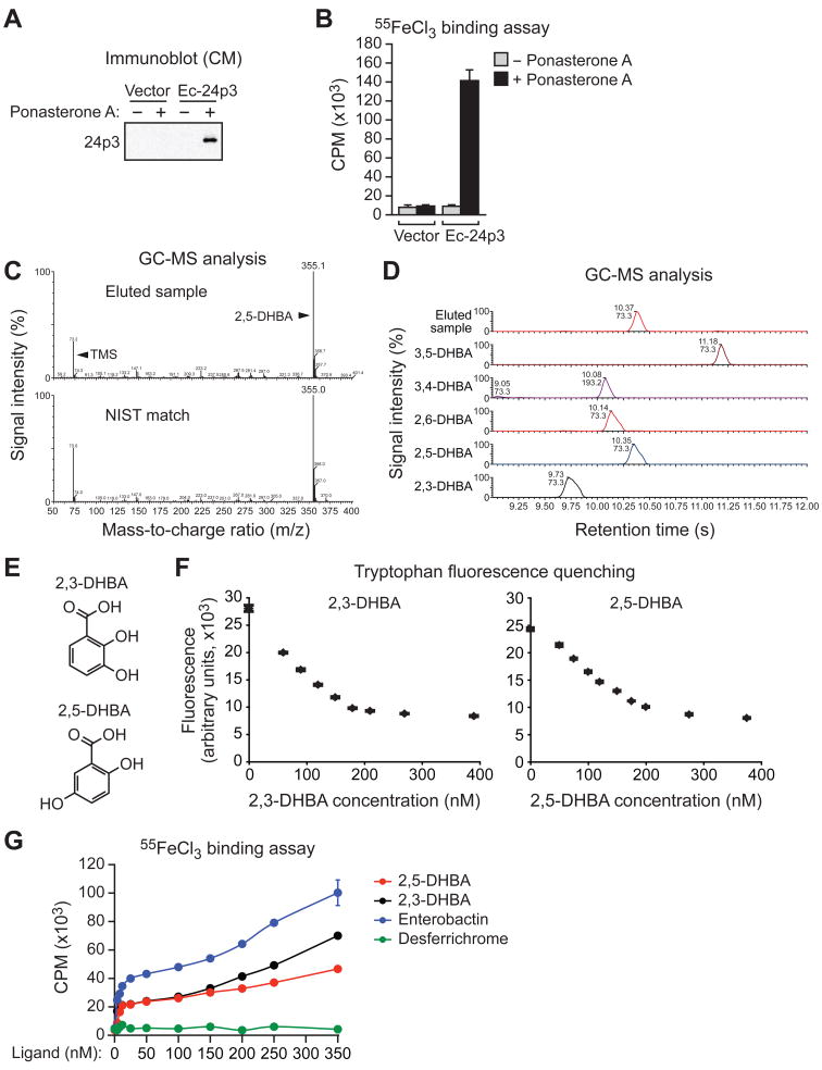
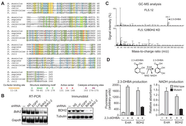
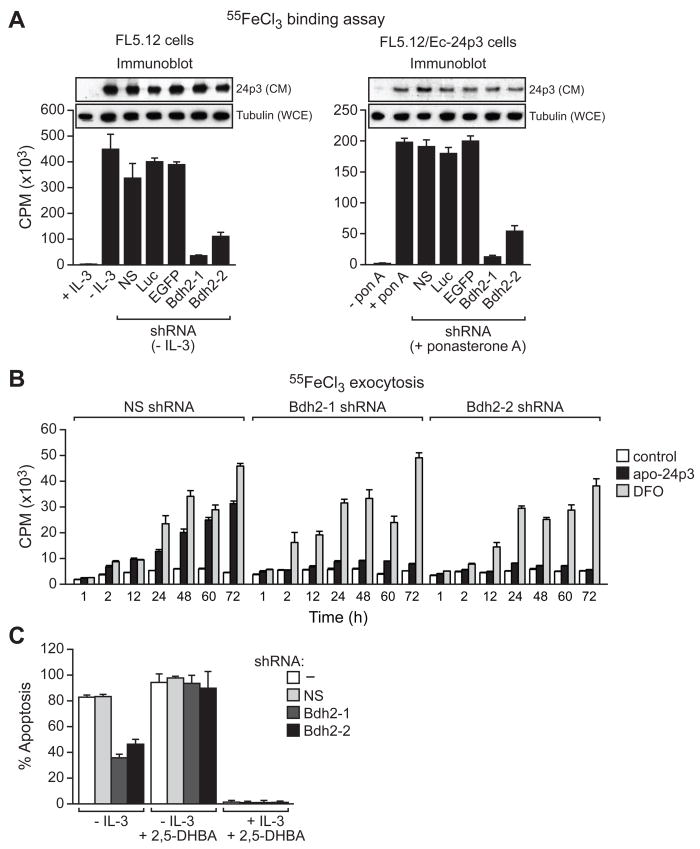
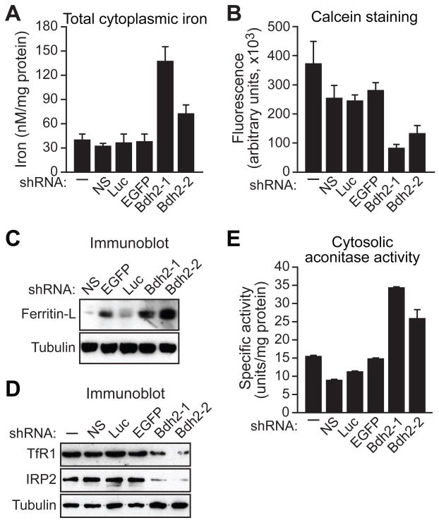
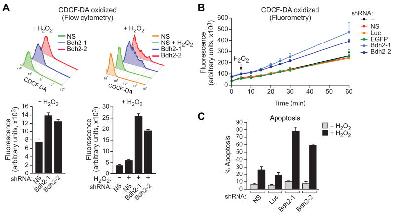
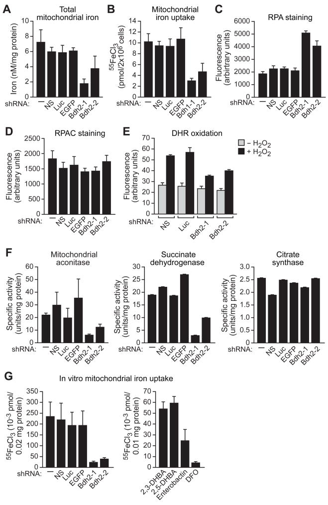
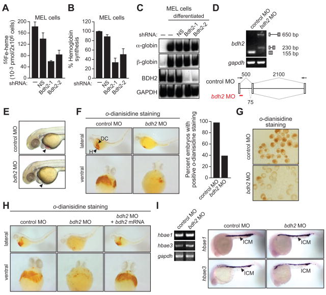
Intracellular iron homeostasis is critical for survival and proliferation. Lipocalin 24p3 is an iron-trafficking protein that binds iron through association with a bacterial siderophore, such as enterobactin, or a postulated mammalian siderophore. Here, we show that the iron-binding moiety of the 24p3-associated mammalian siderophore is 2,5-dihydroxybenzoic acid (2,5-DHBA), which is similar to 2,3-DHBA, the iron-binding component of enterobactin. We find that the murine enzyme responsible for 2,5-DHBA synthesis, BDH2, is the homolog of bacterial EntA, which catalyzes 2,3-DHBA production during enterobactin biosynthesis. RNA interference-mediated knockdown of BDH2 results in siderophore depletion. Mammalian cells lacking the siderophore accumulate abnormally high amounts of cytoplasmic iron, resulting in elevated levels of reactive oxygen species, whereas the mitochondria are iron deficient. Siderophore-depleted mammalian cells and zebrafish embryos fail to synthesize heme, an iron-dependent mitochondrial process. Our results reveal features of intracellular iron homeostasis that are conserved from bacteria through humans.


Figures

Figure 1
Figure 1. 2,5-DHBA is the Iron-Binding Moiety of the 24p3-Associated Mammalian Siderophore
(A) Immunoblot analysis showing 24p3 in the CM of FL5.12 cells expressing either vector or Ec-24p3 in the presence or absence of ponasterone A. (B) Iron binding assay. 24p3 was immunoprecipitated from the CM of cells described in (A) and analyzed for the presence of 55Fe. CPM, counts per minute. Error bars indicate SD. (C) (Top) GC-MS analysis on the TMS-derivatized, small molecule flow-through fraction eluted from immunopurified 24p3. (Bottom) Matching spectrum from the NIST database. (D) GC-MS analysis on the eluted sample from immunopurified 24p3 and a set of DHBA standards. (E) Chemical structures of 2,3-DHBA and 2,5-DHBA. (F) Tryptophan fluorescence quenching assay. 2,3-DHBA (left) or 2,5-DHBA (right) was added to apo-24p3 at increasing concentrations, and tryptophan fluorescence was monitored. Error bars indicate SD. (G) Iron binding assay. Purified apo-24p3 was incubated with 55FeCl3 and increasing concentrations of either 2,3-DHBA, 2,5-DHBA, iron-free enterobactin or desferrichrome, and radioactivity was measured in the 24p3 immunoprecipitate. Error bars indicate SD. See also Figure S1.
Figure 2
Figure 2. Identification of a Murine Homologue of Bacterial EntA that Mediates 2,5-DHBA Synthesis
(A) Multiple sequence alignment of EntA/BDH2 homologues. Identical residues between all five proteins are indicated in yellow; conserved residues in green, and semi-conserved residues in blue. (B) RT-PCR (left) and immunoblot (right) analysis monitoring murine Bdh2 expression in FL5.12 cells treated with one of two unrelated shRNAs targeting Bdh2 or a control non-silencing (NS), enhanced green fluorescent protein (EGFP) or luciferase (Luc) shRNA. (C) GC-MS analysis of the eluted 24p3 sample prepared from parental FL5.12 cells or FL5.12 cells expressing a Bdh2 shRNA. (D) Wild-type and mutant bacterial EntA and mouse BDH2 proteins were analyzed for their ability to convert 2,3-diDHBA to 2,3-DHBA (left) or NAD to NADH (right). Error bars indicate SD. Mutant EntA proteins were expressed at comparable levels to the wild-type proteins (Figure S2C). See also Figure S2.
Figure 3
Figure 3. BDH2 is Required for 24p3-Mediated Iron Transport and Apoptosis
(A) Iron binding assay. (Left) FL5.12 cells were treated in the presence or absence of IL-3, and with a Bdh2 or control shRNA, and the iron-binding ability of 24p3 in the CM was monitored as described in Figure 1B. Inset, immunoblot analysis monitoring 24p3 levels in the CM and tubulin levels in the whole cell extract (WCE). (Right) FL5.12/Ec-24p3 cells were treated in the presence or absence of ponasterone A. Error bars indicate SD. (B) Iron exocytosis assay. 55FeCl3-loaded FL5.12 cells were either untreated (control) or treated with apo-24p3 or DFO, and the presence of 55Fe in the medium was quantified. (C) Apoptosis assay. FL5.12 cells cultured in the presence or absence of IL-3, with or without 2,5-DHBA, and expressing a NS or Bdh2 shRNA were analyzed for apoptosis by annexin V-FITC staining.
Figure 4
Figure 4. BDH2 is Required to Maintain Normal Cytoplasmic Iron Levels
(A–E) FL5.12 cells expressing a Bdh2 or control shRNA were analyzed for total cytoplasmic iron levels by colorimetric analysis (A), for free cytoplasmic iron concentration by fluorescence calcein assay (B), for ferritin-L (C) and Tfr1 and IRP2 (D) levels by immunoblot analysis, and for cytosolic aconitase activity (E). Error bars indicate SD. See also Figures S3A–D and S4.
Figure 5
Figure 5. The Siderophore Protects Cells from Oxidative Stress
(A) FL5.12 cells expressing a NS or Bdh2 shRNA and treated in the presence or absence of hydrogen peroxide (H2O2) were analyzed for ROS by monitoring CDCF-DA fluorescence using flow cytometry. Error bars indicate SD. (B) FL5.12 cells expressing a control or Bdh2 shRNA were analyzed for ROS by monitoring CDCF-DA fluorescence using fluorometry. Error bars indicate SD. (C) FL5.12 cells expressing a control or Bdh2 shRNA were treated in the presence or absence of H2O2 and apoptosis was measured by annexin V-FITC staining. Error bars indicate SD. See also Figure S3E.
Figure 6
Figure 6. BDH2 is Required for Mitochondrial Iron Homeostasis
(A–F) FL5.12 cells expressing a Bdh2 or control shRNA were analyzed for mitochondrial iron levels by colorimetric analysis (A), for mitochondrial iron uptake (B), for RPA (C) and RPAC (D) fluorescence by fluorometry, for DHR 123 oxidation in the presence or absence of H2O2 by flow cytometry (E), and for activities of mitochondrial aconitase, succinate dehydrogenase and citrate synthase (F). (G) In vitro mitochondrial iron import. (Left) Proteolytically treated cytosolic extracts from control or Bdh2 shRNA-treated FL5.12 cells were added to purified mitochondria, and iron import was determined by monitoring mitochondrial incorporation of 55FeCl3. (Right) Purified mitochondria were treated with 2,3-DHBA, 2,5-DHBA, enterobactin or DFO, and iron import was monitored. See also Figures S3F-J and S5.
Figure 7
Figure 7. Requirement of Vertebrate EntA Homologues for Heme Synthesis
(A) MEL cells expressing a NS or Bdh2 shRNA were differentiated. Following labeling with 55Fe, cells were lysed and heme was extracted and analyzed for 55Fe incorporation. Error bars indicate SD. (B) MEL cells were differentiated as described in (A) and following cell lysis, hemoglobin levels were determined by benzidine reaction. Hemoglobin content was expressed relative to that in untreated MEL cells cultured in the absence of DMSO. Error bars indicate SD. (C) MEL cells were differentiated as described in (A) and α- and β-globin mRNA levels monitored by RT-PCR. The levels of Bdh2 and Gapdh were monitored as controls. (D) RT-PCR analysis. The bdh2 MO was injected into one-cell stage embryos and 24 hr later, RNA was extracted and analyzed by RT-PCR. Treatment with the bdh2 MO resulted in retention of the entire intron (upper band) as well as activation of a cryptic splice site that resulted in partial intron retention (lower band). (E) Phenotypic analysis of control MO- and bdh2 MO-injected embryos at 72 hpf. Bdh2-depleted embryos lack hemoglobinized erythrocytes, as evidenced by the absence of red color (arrow). (F) Whole-mount o-dianisidine staining of control MO and bdh2 MO-injected embryos at 72 hpf. Shown are lateral (top) and ventral (bottom) views of the anterior region of embryos. Control MO-injected embryos show positive o-dianisidine staining at the duct of Cuvier (DC) and the heart (H), which is markedly reduced in bdh2 MO-injected embryos. (Right) Quantification of the percent embryos with normal o-dianisidine staining. (G) Blood cells were isolated from control or bdh2 MO-injected embryos and analyzed for hemoglobinization by o-dianisidine staining. (H) Whole-mount o-dianisidine staining of embryos (72 hpf) injected with a control MO or bdh2 MO, or bdh2 MO-treated embryos co-injected with a bdh2 mRNA. (I) RT-PCR (left) and in situ hybridization analysis (right) for hbae1 and hbae3 expression in control and bdh2 MO-injected embryos. See also Figures S4 and S6.

References

    1. Akerstrom B, Flower DR, Salier JP. Lipocalins: unity in diversity. Biochim Biophys Acta. 2000;1482:1–8. - PubMed
    1. Andrews NC. Forging a field: the golden age of iron biology. Blood. 2008;112:219–230. - PMC - PubMed
    1. Blat D, Weiner L, Youdim MB, Fridkin M. A novel iron-chelating derivative of the neuroprotective peptide NAPVSIPQ shows superior antioxidant and antineurodegenerative capabilities. J Med Chem. 2008;51:126–134. - PubMed
    1. Breuer W, Shvartsman M, Cabantchik ZI. Intracellular labile iron. Int J Biochem Cell Biol. 2008;40:350–354. - PubMed
    1. Brownlie A, Donovan A, Pratt SJ, Paw BH, Oates AC, Brugnara C, Witkowska HE, Sassa S, Zon LI. Positional cloning of the zebrafish sauternes gene: a model for congenital sideroblastic anaemia. Nat Genet. 1998;20:244–250. - PubMed

Publication types