Identification of amyloid plaques in retinas from Alzheimer's patients and noninvasive in vivo optical imaging of retinal plaques in a mouse model
- PMID: 20550967
- PMCID: PMC2991559
- DOI: 10.1016/j.neuroimage.2010.06.020
Identification of amyloid plaques in retinas from Alzheimer's patients and noninvasive in vivo optical imaging of retinal plaques in a mouse model
Abstract
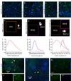
Noninvasive monitoring of β-amyloid (Aβ) plaques, the neuropathological hallmarks of Alzheimer's disease (AD), is critical for AD diagnosis and prognosis. Current visualization of Aβ plaques in brains of live patients and animal models is limited in specificity and resolution. The retina as an extension of the brain presents an appealing target for a live, noninvasive optical imaging of AD if disease pathology is manifested there. We identified retinal Aβ plaques in postmortem eyes from AD patients (n=8) and in suspected early stage cases (n=5), consistent with brain pathology and clinical reports; plaques were undetectable in age-matched non-AD individuals (n=5). In APP(SWE)/PS1(∆E9) transgenic mice (AD-Tg; n=18) but not in non-Tg wt mice (n=10), retinal Aβ plaques were detected following systemic administration of curcumin, a safe plaque-labeling fluorochrome. Moreover, retinal plaques were detectable earlier than in the brain and accumulated with disease progression. An immune-based therapy effective in reducing brain plaques, significantly reduced retinal Aβ plaque burden in immunized versus non-immunized AD mice (n=4 mice per group). In live AD-Tg mice (n=24), systemic administration of curcumin allowed noninvasive optical imaging of retinal Aβ plaques in vivo with high resolution and specificity; plaques were undetectable in non-Tg wt mice (n=11). Our discovery of Aβ specific plaques in retinas from AD patients, and the ability to noninvasively detect individual retinal plaques in live AD mice establish the basis for developing high-resolution optical imaging for early AD diagnosis, prognosis assessment and response to therapies.
Keywords: Alzheimer’s disease; Aβ deposit; Aβ plaque; curcumin; fluorescence; human retina; in vivo optical imaging; mild cognitive impairment; spectral classification; vaccination.
Copyright © 2010 Elsevier Inc. All rights reserved.
Conflict of interest statement
The authors declare that no conflict of interest exists.
Figures


Similar articles
-
Alzheimer's disease in the retina: imaging retinal aβ plaques for early diagnosis and therapy assessment.Neurodegener Dis. 2012;10(1-4):285-93. doi: 10.1159/000335154. Epub 2012 Feb 10. Neurodegener Dis. 2012. PMID: 22343730 Review.
-
Non-invasive optical imaging of retinal Aβ plaques using curcumin loaded polymeric micelles in APPswe/PS1ΔE9 transgenic mice for the diagnosis of Alzheimer's disease.J Mater Chem B. 2020 Aug 26;8(33):7438-7452. doi: 10.1039/d0tb01101k. J Mater Chem B. 2020. PMID: 32662804
-
In vivo Retinal Fluorescence Imaging With Curcumin in an Alzheimer Mouse Model.Front Neurosci. 2020 Jul 3;14:713. doi: 10.3389/fnins.2020.00713. eCollection 2020. Front Neurosci. 2020. PMID: 32719582 Free PMC article.
-
Engineering of donor-acceptor-donor curcumin analogues as near-infrared fluorescent probes for in vivo imaging of amyloid-β species.Theranostics. 2022 Apr 4;12(7):3178-3195. doi: 10.7150/thno.68679. eCollection 2022. Theranostics. 2022. PMID: 35547754 Free PMC article.
-
Retinal Changes in Transgenic Mouse Models of Alzheimer's Disease.Curr Alzheimer Res. 2021;18(2):89-102. doi: 10.2174/1567205018666210414113634. Curr Alzheimer Res. 2021. PMID: 33855942 Review.
Cited by
-
Deciphering the Interacting Mechanisms of Circadian Disruption and Alzheimer's Disease.Neurochem Res. 2021 Jul;46(7):1603-1617. doi: 10.1007/s11064-021-03325-x. Epub 2021 Apr 19. Neurochem Res. 2021. PMID: 33871799 Review.
-
Non-invasive in vivo imaging of brain and retinal microglia in neurodegenerative diseases.Front Cell Neurosci. 2024 Jan 29;18:1355557. doi: 10.3389/fncel.2024.1355557. eCollection 2024. Front Cell Neurosci. 2024. PMID: 38348116 Free PMC article. Review.
-
Systemic administration of Abeta mAb reduces retinal deposition of Abeta and activated complement C3 in age-related macular degeneration mouse model.PLoS One. 2013 Jun 14;8(6):e65518. doi: 10.1371/journal.pone.0065518. Print 2013. PLoS One. 2013. PMID: 23799019 Free PMC article.
-
Biomarkers for Alzheimer's Disease Early Diagnosis.J Pers Med. 2020 Sep 4;10(3):114. doi: 10.3390/jpm10030114. J Pers Med. 2020. PMID: 32899797 Free PMC article. Review.
-
Enhancing foveal avascular zone analysis for Alzheimer's diagnosis with AI segmentation and machine learning using multiple radiomic features.Sci Rep. 2024 Jan 22;14(1):1841. doi: 10.1038/s41598-024-51612-8. Sci Rep. 2024. PMID: 38253722 Free PMC article.
References
-
- Anand P, Kunnumakkara AB, Newman RA, Aggarwal BB. Bioavailability of curcumin: problems and promises. Mol Pharm. 2007;4:807–818. - PubMed
-
- Anderson DH, Talaga KC, Rivest AJ, Barron E, Hageman GS, Johnson LV. Characterization of β amyloid assemblies in drusen: the deposits associated with aging and age-related macular degeneration. Exp Eye Res. 2004;78:243–256. - PubMed
-
- Armstrong RA. β-Amyloid plaques: stages in life history or independent origin? Dement Geriatr Cogn Disord. 1998;9:227–238. - PubMed
-
- Baschong W, Suetterlin R, Laeng RH. Control of autofluorescence of archival formaldehyde-fixed, paraffin-embedded tissue in confocal laser scanning microscopy (CLSM) J Histochem Cytochem. 2001;49:1565–1572. - PubMed
-
- Berisha F, Feke GT, Trempe CL, McMeel JW, Schepens CL. Retinal abnormalities in early Alzheimer's disease. Invest Ophthalmol Vis Sci. 2007;48:2285–2289. - PubMed
Publication types
MeSH terms
Substances
Grants and funding
LinkOut - more resources
Full Text Sources
Other Literature Sources
Medical
Molecular Biology Databases
Miscellaneous

