

Homocysteinethiolactone and paraoxonase: novel markers of diabetic retinopathy
- PMID: 20551012
- PMCID: PMC2928358
- DOI: 10.2337/dc10-0132
Homocysteinethiolactone and paraoxonase: novel markers of diabetic retinopathy
Abstract
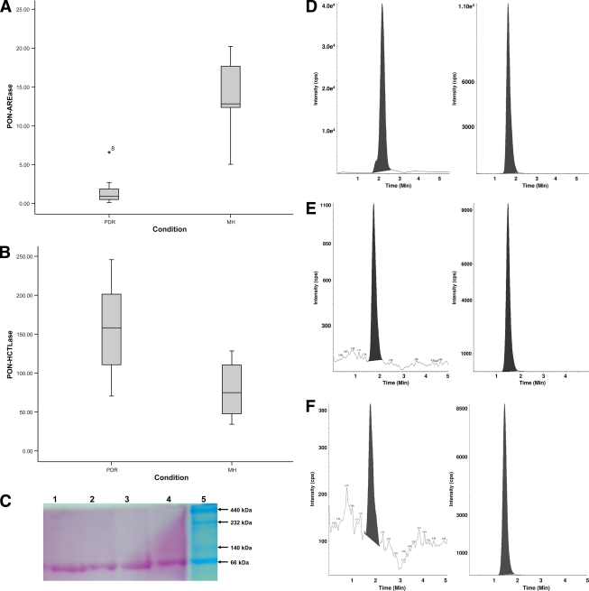
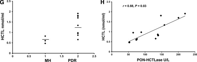
Objective: Paraoxonase (PON) exhibits esterase activity (PON-AREase) and lactonase activity (PON-HCTLase), which prevent LDL oxidation and detoxify homocysteine thiolactone (HCTL). The role of HCTL and PON-HCTLase as a risk factor for the microvascular complication in diabetic retinopathy at the level of vitreous has not been investigated.
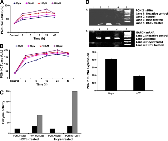
Research design and methods: Undiluted vitreous from patients with proliferative diabetic retinopathy (PDR) (n = 13) and macular hole (MH) (n = 8) was used to determine PON-HCTLase and PON-AREase activity spectrophotometrically. HCTL levels were detected by liquid chromatography-tandem mass spectrometry. In vitro studies were done in primary cultures of bovine retinal capillary endothelial cells (BRECs) to determine the dose- and time-dependent effect of HCTL and homocysteine (Hcys) on PON-HCTLase activity, as well as to determine mRNA expression of PON by RT-PCR.
Results: A significant increase in HCTL and PON-HCTLase activity was observed in PDR compared with MH (P = 0.036, P = 0.001), with a significant positive correlation between them (r = 0.77, P = 0.03). The in vitro studies on BRECs showed a dose- and time-dependent increase in the PON-HCTLase activity and mRNA expression of PON2 when exposed to HCTL and Hcys.
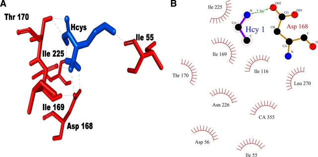
Conclusions: This is the first study showing elevated levels of vitreous HCTL and PON-HCTLase activity in PDR. These elevations are probably a protective effect to eliminate HCTL, which mediates endothelial cell dysfunction. Thus, vitreous levels of HCTL and PON activity can be markers of diabetic retinopathy. The bioinformatics analysis reveals that the structure and function of PON that can be modulated by hyperhomocysteinemia in PDR can affect the dual-enzyme activity of PON.
Figures
References
-
- Clarke R, Daly L, Robinson K, Naughten E, Cahalane S, Fowler B, Graham I: Hyperhomocysteinemia: an independent risk factor for vascular disease. N Engl J Med 1991;324:1149–1155 - PubMed
-
- Gu W, Lu J, Yang G, Dou J, Mu Y, Meng J, Pan C: Plasma homocysteine thiolactone associated with risk of macrovasculopathy in Chinese patients with type 2 diabetes mellitus. Adv Ther 2008;25:914–924 - PubMed
-
- Jakubowski H, Zhang L, Bardeguez A, Aviv A: Homocysteine thiolactone and protein homocysteinylation in human endothelial cells: implications for atherosclerosis. Circ Res 2000;87:45–51 - PubMed
-
- Domagała TB, Łacinski M, Trzeciak WH, Mackness B, Mackness MI, Jakubowski H: The correlation of homocysteine-thiolactonase activity of the paraoxonase (PON1) protein with coronary heart disease status. Cell Mol Biol 2006;52:4–10 - PubMed
-
- Reddy ST, Wadleigh DJ, Grijalva V, Ng C, Hama S, Gangopadhyay A, Shih DM, Lusis AJ, Navab M, Fogelman AM: Human paraoxonase-3 is an HDL-associated enzyme with biological activity similar to paraoxonase-1 protein but is not regulated by oxidized lipids. Arterioscler Thromb Vasc Biol 2001;21:542–547 - PubMed
Publication types
MeSH terms
Substances
LinkOut - more resources
Full Text Sources
Medical
Miscellaneous
