. 2010 Jun 15;24(12):1236-41.
doi: 10.1101/gad.1920310.

Myc protein is stabilized by suppression of a novel E3 ligase complex in cancer cells

Affiliations

Myc protein is stabilized by suppression of a novel E3 ligase complex in cancer cells

Seung H Choi et al. Genes Dev. .

Abstract

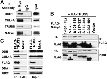
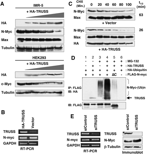
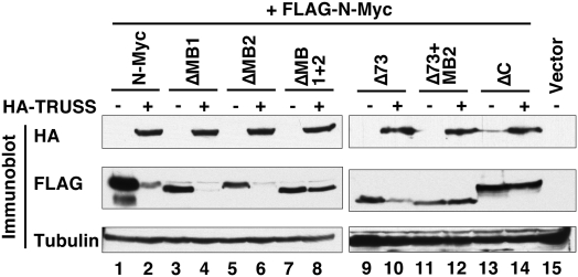
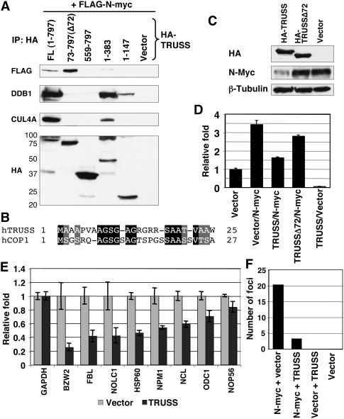
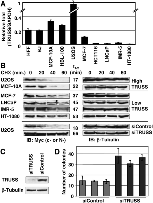
Rapid Myc protein turnover is critical for maintaining basal levels of Myc activity in normal cells and a prompt response to changing growth signals. We characterize a new Myc-interacting factor, TRPC4AP (transient receptor potential cation channel, subfamily C, member 4-associated protein)/TRUSS (tumor necrosis factor receptor-associated ubiquitous scaffolding and signaling protein), which is the receptor for a DDB1 (damage-specific DNA-binding protein 1)-CUL4 (Cullin 4) E3 ligase complex for selective Myc degradation through the proteasome. TRPC4AP/TRUSS binds specifically to the Myc C terminus and promotes its ubiquitination and destruction through the recognition of evolutionarily conserved domains in the Myc N terminus. TRPC4AP/TRUSS suppresses Myc-mediated transactivation and transformation in a dose-dependent manner. Finally, we found that TRPC4AP/TRUSS expression is strongly down-regulated in most cancer cell lines, leading to Myc protein stabilization. These studies identify a novel pathway targeting Myc degradation that is suppressed in cancer cells.


Figures

Figure 1.
Figure 1.
Myc interacts with TRUSS and recruits the DDB1–CUL4 E3 ligase complex. (A) TRUSS forms a complex with native N-Myc. Endogenous N-Myc protein was immunoprecipitated from IMR-5 neuroblastoma cells with an anti-N-Myc monoclonal antibody, B8.4.B. Precipitating proteins were resolved by SDS-PAGE and immunoblotted with the indicated antibodies. Inputs for immunoprecipitations are shown at the right. (B) Coimmunoprecipitation assay to locate the TRUSS-interacting region in N-Myc. HA-tagged full-length TRUSS-expressing vector (HA-TRUSS) was cotransfected with Flag-tagged N-Myc-expressing vectors containing the indicated deletions in N-Myc. Flag-N-Myc immunoprecipitates, together with inputs (5%), were probed for either HA (TRUSS) or Flag (N-Myc). (C) TRUSS associates with the DDB1–CUL4A complex. TRUSS was immunoprecipitated and immunoblotted from HeLa cells that stably expressed Flag-TRUSS. Antibodies used for immunoblots are described on the left. The input was split for each immunoprecipitation.
Figure 2.
Figure 2.
TRUSS enhances Myc protein turnover. (A) TRUSS was expressed ectopically in IMR-5 cells (top panel) and HEK293 cells (bottom panel) at increasing dosages (0, 0.2, 0.5, 1, 2, and 3 μg, respectively, supplemented with an empty vector for constant DNA input). Total cell lysates were prepared after 36 h, and equal amounts of protein were loaded for immunoblot with indicated antibodies to detect native Myc protein levels. β-Tubulin was used as a loading control. (B) TRUSS does not affect Myc mRNA levels. TRUSS was expressed ectopically in IMR-5 cells (3 μg DNA), and total RNA was isolated and subjected to RT–PCR for N-myc mRNA after 36 h. GAPDH served as an input control. (C) Ectopic TRUSS expression shortens N-Myc protein half-life in IMR-5 cells. TRUSS or empty vector was expressed ectopically for 24 h, then cycloheximide (50 μg/mL) was added to the media to inhibit de novo protein synthesis. Whole-cell lysates were resolved by SDS-PAGE followed by immunoblotting. Max antibody was used as an input control. (D) TRUSS promotes N-Myc ubiquitination. Flag-N-myc, HA-TRUSS, and HA-Ubiquitin were cotransfected in HEK 293 cells ± MG-132. Flag-N-mycΔC was transfected in lane 5 instead of wild-type N-myc. N-Myc was immunoprecipitated with anti-Flag beads, and was immunoblotted with anti-HA antibody to detect TRUSS and high-molecular ubiquitin-conjugated N-Myc. In the absence of MG-132, N-Myc protein is degraded without accumulating ubiquitinated products. (E) Depletion of TRUSS leads to elevated N-Myc protein levels but no change in mRNA. siRNA against human TRUSS and control siRNA were transfected to IMR-5 cells to knock down TRUSS expression. (Left panel) After 36 h, RT–PCR was performed to evaluate the efficiency of siRNA. (Right panel) At the same time, total cell lysates were harvested to measure protein levels of TRUSS and N-Myc. β-Tubulin served as a loading control.
Figure 3.
Figure 3.
TRUSS requires both N-terminal and C-terminal Myc domains for degradation. Full-length N-myc and various deletion mutants were expressed in HEK 293 cells in the absence or presence of TRUSS coexpression to map the TRUSS-dependent Myc degrons. Total lysates were immunoblotted to compare the net amount of Myc protein. ΔMB1, ΔMB2, and ΔC correspond to deletion of N-Myc amino acids 40–58, 103–119, and 372–454, respectively.
Figure 4.
Figure 4.
The TRUSS N terminus is necessary for recruiting the DDB1–CUL4 complex and repressing transcription of Myc target genes. (A) HA-tagged full-length TRUSS or mutants containing different deletions were coexpressed with Flag-tagged N-myc. Expressed regions of TRUSS are indicated at the top. Immune complexes were resolved by SDS-PAGE and were blotted with the indicated antibodies. (B) Alignment between N-terminal regions of TRUSS and hCOP1. (C) Full-length and an N-terminal deletion (Δ72) of TRUSS were expressed in IMR-5 cells. Total-cell lysates were harvested and analyzed by immunoblotting. β-Tubulin is shown as a loading control. (D) Reporter construct harboring Myc-binding sites was analyzed in U2OS cells. Assays were performed 24 h after transfection. Luciferase activities were normalized to Renilla reporter activity as an internal control. Values were normalized to the vector control as 1. (E) RT–PCR analysis of Myc target genes in IMR-5 cells. A TRUSS expression vector was transfected to IMR-5 cells, and total RNAs were isolated for RT–PCR 36 h after transfection. GAPDH was used as an input control. (F) RK3E foci assay was carried out to measure the oncogenic potential of Myc in response to TRUSS. The numbers of foci from three independent transfections were averaged.
Figure 5.
Figure 5.
Down-regulation of TRUSS expression correlates with prolonged Myc half-life in cancer cells. (A) Total RNA from the indicated cell lines was analyzed by RT–PCR. TRUSS expression was normalized to GAPDH to compare the relative expression between lines. Data are the average of three independent assays. (B, top and middle) Half-life of Myc proteins from different human cell lines used in A are shown. Cycloheximide (CHX, 50 μg/mL) was added to the media, and whole-cell lysates were resolved by SDS-PAGE and were immunoblotted with anti-c-Myc antibody (anti-N-Myc antibody was used for IMR-5). After detection of Myc protein, the same blots were reprobed with anti-β-Tubulin antibody for a loading control. Myc half-lives calculated from image analysis are shown (t1/2). (Bottom) Knockdown of TRUSS expression prolongs c-Myc half-life in U2OS cells. c-Myc half-lives with t1/2 of 18 min in control cells and extended to 66 min after TRUSS knockdown. β-Tubulin served as a loading control. (C) Knockdown of TRUSS mRNA by siRNA in U2OS cells. (D) U2OS cells are embedded in soft-agar medium after siRNA transfection against TRUSS. After 20 d, colonies >150 μm are counted from three different areas. Three independent transfections were carried out.

Comment in

References

    1. Bazarov AV, Adachi S, Li SF, Mateyak MK, Wei S, Sedivy JM 2001. A modest reduction in c-myc expression has minimal effects on cell growth and apoptosis but dramatically reduces susceptibility to Ras and Raf transformation. Cancer Res 61: 1178–1186 - PubMed
    1. Bonvini P, Nguyen P, Trepel J, Neckers LM 1998. In vivo degradation of N-myc in neuroblastoma cells is mediated by the 26S proteasome. Oncogene 16: 1131–1139 - PubMed
    1. Chang DW, Claassen GF, Hann SR, Cole MD 2000. The c-Myc transactivation domain is a direct modulator of apoptotic versus proliferative signals. Mol Cell Biol 20: 4309–4319 - PMC - PubMed
    1. Cohn SL, Salwen H, Quasney MW, Ikegaki N, Cowan JM, Herst CV, Kennett RH, Rosen ST, DiGiuseppe JA, Brodeur GM 1990. Prolonged N-myc protein half-life in a neuroblastoma cell line lacking N-myc amplification. Oncogene 5: 1821–1827 - PubMed
    1. Cowling VH, Cole MD 2006. Mechanism of transcriptional activation by the Myc oncoproteins. Semin Cancer Biol 16: 242–252 - PubMed

Publication types