. 2010 Jun 15;24(12):1266-80.
doi: 10.1101/gad.571710.

Functional conservation of Mei4 for meiotic DNA double-strand break formation from yeasts to mice

Affiliations

Functional conservation of Mei4 for meiotic DNA double-strand break formation from yeasts to mice

Rajeev Kumar et al. Genes Dev. .

Abstract

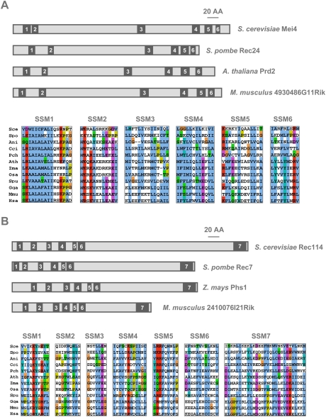
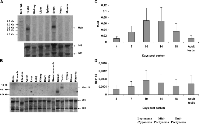
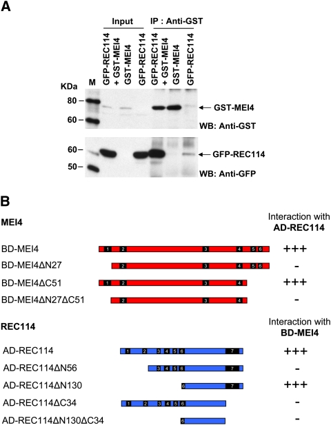
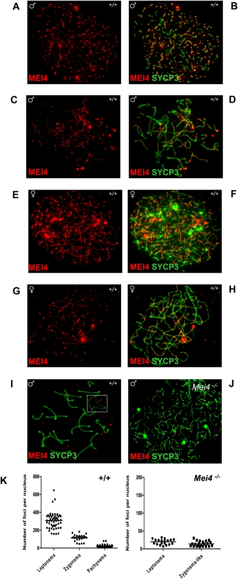
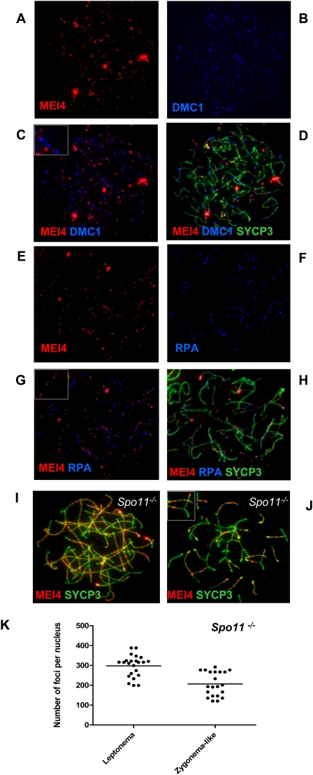
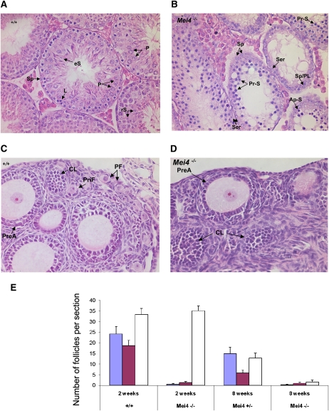
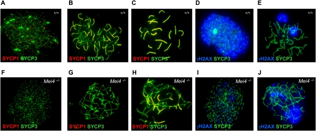
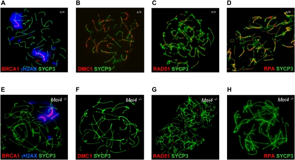
Meiotic recombination is initiated by the programmed induction of DNA double-strand breaks (DSBs) catalyzed by the evolutionarily conserved Spo11 protein. Studies in yeast have shown that DSB formation requires several other proteins, the role and conservation of which remain unknown. Here we show that two of these Saccharomyces cerevisiae proteins, Mei4 and Rec114, are evolutionarily conserved in most eukaryotes. Mei4(-/-) mice are deficient in meiotic DSB formation, thus showing the functional conservation of Mei4 in mice. Cytological analyses reveal that, in mice, MEI4 is localized in discrete foci on the axes of meiotic chromosomes that do not overlap with DMC1 and RPA foci. We thus propose that MEI4 acts as a structural component of the DSB machinery that ensures meiotic DSB formation on chromosome axes. We show that mouse MEI4 and REC114 proteins interact directly, and we identify conserved motifs as required for this interaction. Finally, the unexpected, concomitant absence of Mei4 and Rec114, as well as of Mnd1, Hop2, and Dmc1, in some eukaryotic species (particularly Neurospora crassa, Drosophila melanogaster, and Caenorhabditis elegans) suggests the existence of Mei4-Rec114-dependent and Mei4-Rec114-independent mechanisms for DSB formation, and a functional relationship between the chromosome axis and DSB formation.


Figures

Figure 1.
Figure 1.
Evolutionary conservation of Mei4 and Rec114 in eukaryotes. The relative localization of the conserved SSMs within the primary structures of fungal, plant, and mouse Mei4 (A) or Rec114 (B) orthologs are shown in the top panels. Alignments of the corresponding SSMs from the same and other representative eukaryotes are shown in the bottom panels. Conserved sequence blocks were extracted from multiple protein sequence alignments generated by MAFFT and were colored with Jalview using default ClustalX color schemes (using the MPI bioinformatics Web resources; see the Materials and Methods). Except for glycines and prolines, colors (i.e., WLVIMFAC in blue, KR in red, TSNQ in green, DE in magenta, G in orange, HY in cyan, and P in yellow) were assigned to residues if the amino acid profile at the given position was conserved. (Sce) Saccharomyces cerevisiae; (Spo) Schizosaccharomyces pombe; (Ani) Aspergillus nidulans; (Cci) Coprinopsis cinerea; (Pch) Phanerochaete chrysosporium; (Ath) Arabidopsis thaliana; (Zma) Zea mays; (Osa) Oryza sativa; (Spu) Strongylocentrotus purpuratus; (Gga) Gallus gallus; (Mmu) Mus musculus; (Hsa) Homo sapiens sapiens.
Figure 2.
Figure 2.
Mei4 and Rec114 are expressed in testis and during the first wave of spermatogenesis. (A,B) Northern blot hybridization of RNA extracted from various tissues of adult mice with the Mei4 (A) or Rec114 (B) probe. Bottom panels show rRNAs (18S and 28S) on the membranes stained with methylene blue. (C,D) RT-qPCR amplification of RNA extracted from testes of juvenile and adult mice. For each gene, qPCR was performed with two different primer pairs specific for Mei4 (C) or Rec114 (D). Plotted values are the expression levels normalized to β-Actin (means of five qPCR assays with 95% confidence interval).
Figure 3.
Figure 3.
MEI4 interacts with REC114. (A) Coimmunoprecipitation of MEI4 and REC114. HeLa cells were transfected with plasmids expressing mouse GST-MEI4 and/or GFP-REC114. Protein extracts were immunoprecipitated with anti-GST or anti-GFP (Supplemental Fig. S5A) antibodies. Inputs (1% of total) and immunoprecipitated fractions (40% of total) were detected with anti-GST or anti-GFP antibodies. Protein standards were used as molecular weight markers (M). (B) Mapping the MEI4 and REC114 interaction domains by yeast two-hybrid assay. The ability to interact of full-length and N-terminal or C-terminal deletion mutants of MEI4 and REC114 was assessed in a yeast two-hybrid assay. Either strong interaction (+++) or no interaction (−) was observed, based on growth on selective media (see also Supplemental Fig. S6).
Figure 4.
Figure 4.
Discrete MEI4 foci on chromosome axes of spermatocytes at leptonema and zygonema. MEI4 was detected in leptotene or zygotene spermatocytes (A–D) and oocytes at E16 (E–H). MEI4 was not observed on the axes of autosomes and sex chromosomes (white rectangle) at pachynema (I), and in spermatocytes from Mei4−/− mice (J). (K) Foci revealed by the anti-MEI4 antibody were quantified in wild-type at leptonema (mean = 309 foci per nucleus, n = 51), zygonema (mean = 114, n = 28), and pachynema (mean = 24, n = 36). Counts included all nuclear foci, of which 67%, 49%, and 14% were on axes at leptonema, zygonema, and pachynema, respectively. On average, 20 and 14 foci per nucleus were detected in the control experiment using Mei4−/− leptotene and zygotene-like nuclei, of which 20% and 23% were axis-associated, respectively. The anti-SYCP3 antibody was used to detect axial elements. (A,C,E,G:) Anti-MEI4 antibody alone. (B,D,F,H,I,J) Anti-MEI4 and anti-SYCP3 antibodies.
Figure 5.
Figure 5.
MEI4 foci do not colocalize with DMC1 and RPA, and do not require SPO11. MEI4 was detected in parallel with DMC1 (A–D) or RPA (E–H). Insets in C and G show enlarged views of MEI4, DMC1, and RPA foci. MEI4 foci were detected and quantified in Spo11/ spermatocytes. On average, 298 and 207 foci per nucleus were detected at leptonema (n = 23) and zygonema-like (n = 21), respectively (I–K). Inset in J shows the absence of MEI4 in a synapsed region.
Figure 6.
Figure 6.
Meiotic defects in testis and ovary from Mei4−/− mice. (A,B) Hematoxylin-eosin staining of testis from adult wild-type (A) and Mei4−/− (B) mice (n = 2 for each genotype). Among primary spermatocytes, some stages observed in wild-type testis were not present in Mei4−/− (i.e., pachynema, diplonema), whereas spermatocytes with apoptotic nuclei were frequent. Haploid cells (spermatids and spermatozoa) were never observed in Mei4−/−. (Sp) Spermatogonia; (eS) elongated spermatids; (rS) round spermatids; (P) pachynema; (Z) zygonema; (L) leptonema; (Ser) Sertoli; (Pr-S) primary spermatocyte at leptonema or zygonema; (PL) preleptonema; (Ap-S) apoptotic nuclei. (C,D) Hematoxylin-eosin staining of ovaries from 2-wk-old wild-type (C) or Mei4−/− (D) mice shows alteration in oogenesis. (PF) Primordial follicle; (CL) corpus luteum; (PreA) preantral oocyte; (PriF) primary follicle. (E) Quantification of primordial follicles (blue), primary follicles (purple), and growing follicles (white) in 2-wk-old (n = 3 for each genotype) and 8-wk-old (n = 1 for each genotype) wild-type, Mei4+/−, and Mei4−/− ovaries (error bars indicate 95% confidence intervals).
Figure 7.
Figure 7.
Mei4 is required for DSB and homologous synapsis formation. Spreads of wild-type (A–E) and Mei4−/− (F–J) spermatocytes (n = 3 for each genotype) were analyzed with antibodies directed against SYCP1 (which is used to monitor synapsis formation) and SYCP3 (A,B,C,F,G,H), and γH2AX (a marker of DSB formation) and SYCP3 (D,E,I,J).
Figure 8.
Figure 8.
Mei4 is required for sex body formation and loading of the DSB repair proteins DMC1, RAD51, and RPA. Spreads of wild-type (A–D) and Mei4−/− (E–H) spermatocytes (n = 3 for each genotype) were analyzed with antibodies directed against BRCA1/γH2AX/SYCP3 (A,E), DMC1/SYCP3 (B,F), RAD51/SYCP3 (C,G), and RPA/SYCP3 (D,H).

Comment in

References

    1. Arora C, Kee K, Maleki S, Keeney S 2004. Antiviral protein ski8 is a direct partner of spo11 in meiotic DNA break formation, independent of its cytoplasmic role in RNA metabolism. Mol Cell 13: 549–559 - PubMed
    1. Barchi M, Mahadevaiah S, Di Giacomo M, Baudat F, de Rooij DG, Burgoyne PS, Jasin M, Keeney S 2005. Surveillance of different recombination defects in mouse spermatocytes yields distinct responses despite elimination at an identical developmental stage. Mol Cell Biol 25: 7203–7215 - PMC - PubMed
    1. Baudat F, de Massy B 2007. Regulating double-stranded DNA break repair towards crossover or non-crossover during mammalian meiosis. Chromosome Res 15: 565–577 - PubMed
    1. Baudat F, Manova K, Yuen JP, Jasin M, Keeney S 2000. Chromosome synapsis defects and sexually dimorphic meiotic progression in mice lacking spo11. Mol Cell 6: 989–998 - PubMed
    1. Bellani MA, Romanienko PJ, Cairatti DA, Camerini-Otero RD 2005. SPO11 is required for sex-body formation, and Spo11 heterozygosity rescues the prophase arrest of Atm−/− spermatocytes. J Cell Sci 118: 3233–3245 - PubMed

Publication types

MeSH terms