. 2010 Jun 15;24(12):1295-305.
doi: 10.1101/gad.1921510.

Cdx2 regulates endo-lysosomal function and epithelial cell polarity

Affiliations

Cdx2 regulates endo-lysosomal function and epithelial cell polarity

Nan Gao et al. Genes Dev. .

Abstract

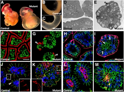
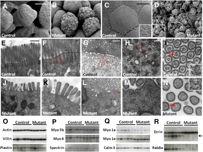
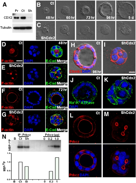
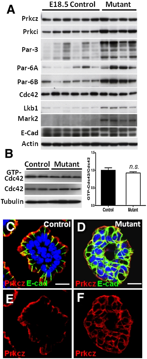
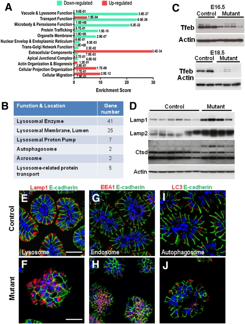
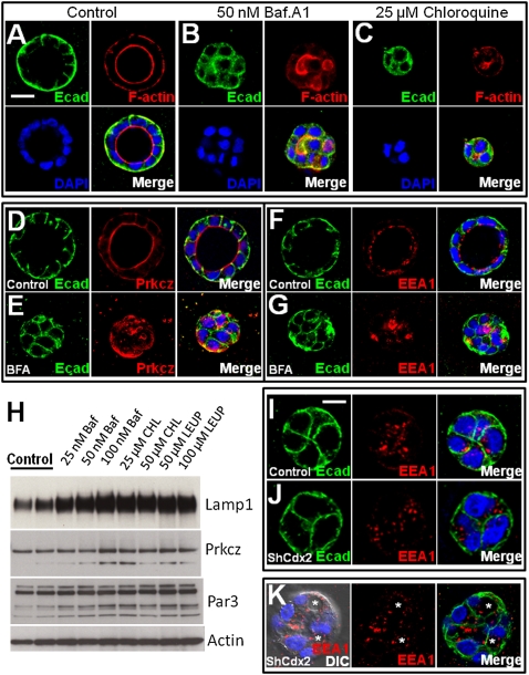
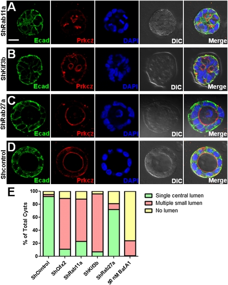
In contrast to our significant understanding of signaling cascades that determine cell polarity in lower eukaryotic or immortalized cells, little is known about the transcriptional program that governs mammalian epithelial polarization in vivo. Here we show, using conditional gene ablation and three-dimensional tissue culture, that the homeobox transcription factor Cdx2 controls apical-basolateral polarity in mouse enterocytes and human colonic epithelial cells. Cdx2 regulates a comprehensive gene network involved in endo-lysosomal maturation and protein transport. In the absence of Cdx2, defective protein trafficking impairs apical-basal transport and induces ectopic lumen formation. These defects are partially recapitulated by suppression of key apical transport components, Rab11a and Kif3b, which are regulated by Cdx2. Furthermore, Cdx2 deficiency affects components that control the organization of microvillus actin cytoskeleton, leading to severe microvillus atrophy. These results demonstrate that Cdx2 regulates epithelial cell polarity and morphogenesis through control of apical protein transport and endo-lysosomal function.


Figures

Figure 1.
Figure 1.
Intestine-specific ablation of Cdx2 causes altered enterocyte morphology and apical/basolateral polarity. (A–C) E18.5 gastrointestinal tracts, examined by light microscopy, revealed translucent appearance of the Cdx2 mutant intestinal tube. Matched control and mutant (Cdx2loxP/loxP; VillinCre) duodenal segments are shown in B and C at higher magnification. (D,–E) TEM micrographs of E18.5 control and Cdx2 mutant intestinal villus cross-sections. The arrow in E points to one of the enlarged vacuoles in the mutant cells. (F,G) The brush border marker alkaline phosphatase was immunostained and is visualized in red. Nuclei are labeled in green. Arrows point to the abnormal distribution of alkaline phosphatase activity on the entire Cdx2 mutant cell membrane. (H,I) Costaining of DBA lectin (red) and E-cadherin (green). Arrows point to the inclusion of lectin staining within subapical cytoplasm in mutant cells. (J,K) Costaining of ZO-1 tight junction protein (red) and E-cadherin (green). The arrow points to ZO-1 localization at tight junctions in control cells. (L,M) Costaining of the basement membrane marker laminin (red) and E-cadherin (green). Establishment of the basement membrane is clearly perturbed in the absence of Cdx2. Nuclei were labeled by DAPI in H–M. Bars: D,E, 10 μm; F–M, 20 μm.
Figure 2.
Figure 2.
Intestine-specific Cdx2 deficiency results in abnormal microvillus morphogenesis. (A–D) SEM demonstrates “uneven” luminal cell surface and abnormal microvillus morphology in Cdx2 mutant intestines. (E,F,J,K) Vertical TEM sections show shortened microvilli in Cdx2 mutant cells (shown in J). In addition, the normal bundled appearance of actin filaments at the microvillar core is absent in Cdx2-deficient cells (K), as compared with control cells (E,F). Red dotted lines mark two bundled actin filaments extending from the microvilli into the apical cytoplasm. (G,H,L,M) Transverse TEM sections show that actin microfilament bundles, indicated by red broken circles in control cellular terminal web in G and H, are missing in mutant cells (shown in L, M). Mutant cells develop canaliculi marked by red dotted lines in L. (I,N) Transverse TEM section of microvilli reveals absence of bundled actin filaments at the microvillar core in Cdx2 mutant cells (shown in N). Red arrows point to the core actin filaments. (O–R) Western blots for protein components of the microvillar core actin filaments (O), the cytoplasmic terminal web (P), the bridging proteins between the actin filaments and the plasma membrane, and Rab8a, the small GTPase regulating apical protein transport (Q,R). Arrow in R points to the smaller products, which were present only in control intestines, recognized by Ezrin antibody. Bars: A,B,G,L, 50 μm; C,D, 5 μm; E,J, 500 nm; F,H,I,K,M,N, 100 nm.
Figure 3.
Figure 3.
CDX2 is required for apical–basal polarity in human Caco-2 cells. (A) Western blot for CDX2 demonstrates efficient knockdown of CDX2 expression in lentiviral shCDX2 transduced Caco-2 cells (Sh), compared with parental cells (Pr) and nontargeting lentiviral particle-treated control cells (Ct), at first passage after viral transduction. (B,C) Control Caco-2 cells form cysts in 3D culture within 72 h, demonstrating a central lumen, while CDX2 knockdown cells fail to form a primary lumen even after 5-d culture. (D–G) Cysts were stained for F-actin (red), E-cadherin (green), and DAPI (blue) at various time points. Note that CDX2 knockdown cells elaborate multiple small lumens at 48 h (E), but fail to promote a central lumen (G). (H,I) Cysts were stained for basolateral marker CTNNB1 (β-catenin) (red). (J,K) Cysts were stained for a basal transporter, Na+/K+-ATPase (green). (L,M) Control and CDX2-deficient Caco-2 cysts were stained for PRKCZ in red. Nuclei were labeled by DAPI in blue. (N) In vitro protein kinase C assay demonstrates a reduction of PRKCZ kinase activity in CDX2-deficient cells by 25.6%. Bars, 20 μm.
Figure 4.
Figure 4.
Cdx2 deficiency leads to elevated expression of Par components, and mislocalization of Prkcz. (A) Intestine-specific Cdx2 deficiency leads to increased levels of several of Par polarity proteins, but not Cdc42. (B) Total or activated Cdc42 levels are not changed in Cdx2 mutant intestines. (C–F) Immunofluorescent staining of Prkcz (red) and E-cadherin (green) demonstrates nonapical localization of Prkcz localization in mutant cells. Nuclei were labeled by DAPI in C and D. Bars: 12.87 μm.
Figure 5.
Figure 5.
Cdx2 deficiency impairs endo-lysosomal maturation and function in enterocytes. (A) Using high-stringency gene classification, functional annotation of the expression profile of normal and mutant E18.5 intestines reveals that the gene category of “vacuole and lysosome” has the highest enrichment score among the down-regulated genes, followed by “transport function,” “microbody and peroxisome,” “protein trafficking,” and “organelle membrane.” The statistical significance of the enrichment of each category is indicated as the P-value on the side of the corresponding bar. (B) Eighty-two endo-lysosomal genes involving various aspects of endosomal and lysosomal biology are reduced in mutant intestines. (C) Tfeb, a transcription factor identified by microarray analysis, is initially reduced in the E16.5, and is decreased further in E18.5 Cdx2 mutant intestines. (D) Western blots for lysosomal markers (Lamp1, Lamp2, and Ctsd) using control and Cdx2 mutant intestinal lysates. (E,F) Lamp1 staining (red) in control and Cdx2 mutant enterocytes. (G,H) Immunofluerescent staining for the early endosome marker EEA1 (red). (I,J) Immunofluorescent staining for the autophagosome marker LC3 (red). Basolateral plasma membranes were labeled with E-cadherin (green). Nuclei were labeled by DAPI. Bars: E–J, 12 μm.
Figure 6.
Figure 6.
Blockage of endo-lysosomal maturation perturbs apical–basal cell polarity. (A–C) Caco-2 3D cyst formation assays were performed with 3-d incubation with normal growth medium (control), or medium containing 50 nM Baf.A1 or 25 μM CHL. F-actin and E-cadherin were stained in red and green, respectively. (D,E) Control and Baf.A1-treated cysts were stained for Prkcz (red) and E-cadherin (green). (F,G) Control and Baf.A1-treated cysts were stained for EEA1 (red) and E-cadherin (green). (H) Western blots for Lamp1, Prkcz, and Par3 using lysates from Caco-2 cells treated with inhibitors overnight at indicated concentrations. Leupeptin serves as a nonspecific protease inhibitor. β-Actin serves as a loading control. (I,J) Early cysts from control and CDX2 knockdown cells were stained for EEA1 (red) and E-cadherin (green). Note that control endosomes are clustered apically into the center where the prospective lumen will emerge. (K) Three-day-old CDX2 knockdown cysts were stained with EEA1 (red) and E-cadherin (green). Note that the knockdown cells are able to accumulate endosomes around several ectopic lumens (asterisks). Bars: A–G,K, 20 μm; I,J, 9 μm.
Figure 7.
Figure 7.
Suppression of apical transport proteins recapitulates CDX2 knockdown polarity defects. (A–D) Caco-2 cysts derived from RAB11A, KIF3B, and RAB27A knockdown cells, as well as control cells infected with nontargeting shRNA viral particles, were stained for PRKCZ (red) and E-cadherin (green). Bar, 20 μm. (E) Percentage of cysts with single central lumen, no lumen, or multiple lumens were quantified. Data represent multiple independent experiments.

Similar articles

Cited by

References

    1. Ameen N, Apodaca G 2007. Defective CFTR apical endocytosis and enterocyte brush border in myosin VI-deficient mice. Traffic 8: 998–1006 - PubMed
    1. Baricault L, Garcia M, Cibert C, Sapin C, Geraud G, Codogno P, Trugnan G 1993. Forskolin blocks the apical expression of dipeptidyl peptidase IV in Caco-2 cells and induces its retention in lamp-1-containing vesicles. Exp Cell Res 209: 277–287 - PubMed
    1. Barth AI, Caro-Gonzalez HY, Nelson WJ 2008. Role of adenomatous polyposis coli (APC) and microtubules in directional cell migration and neuronal polarization. Semin Cell Dev Biol 19: 245–251 - PMC - PubMed
    1. Cramm-Behrens CI, Dienst M, Jacob R 2008. Apical cargo traverses endosomal compartments on the passage to the cell surface. Traffic 9: 2206–2220 - PubMed
    1. Debnath J, Muthuswamy SK, Brugge JS 2003. Morphogenesis and oncogenesis of MCF-10A mammary epithelial acini grown in three-dimensional basement membrane cultures. Methods 30: 256–268 - PubMed

Publication types

MeSH terms