. 2010 Jun 15;24(12):1306-16.
doi: 10.1101/gad.570310.

A novel phosphatase cascade regulates differentiation in Trypanosoma brucei via a glycosomal signaling pathway

Affiliations

A novel phosphatase cascade regulates differentiation in Trypanosoma brucei via a glycosomal signaling pathway

Balázs Szöor et al. Genes Dev. .

Abstract

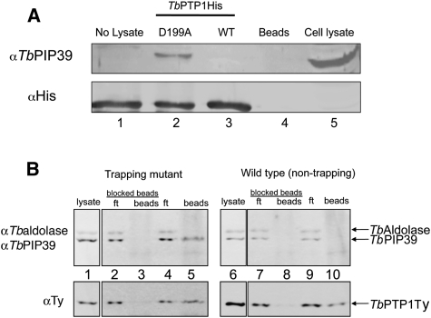
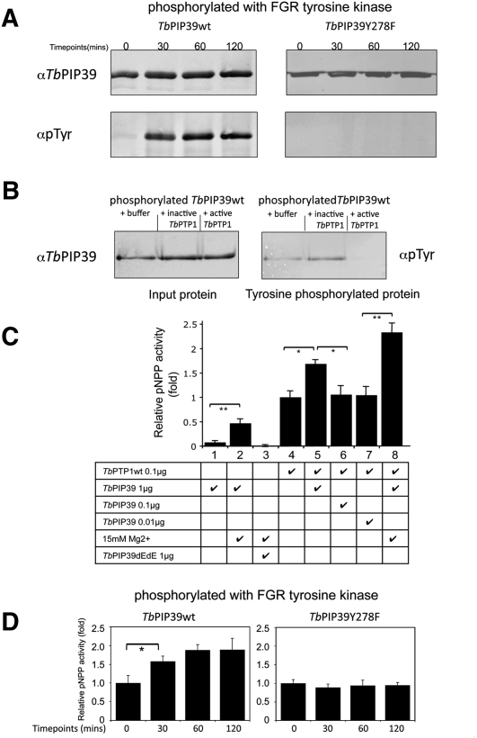
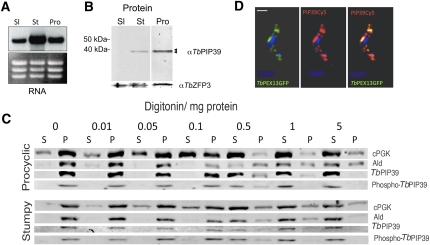
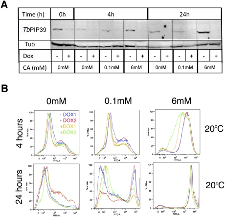
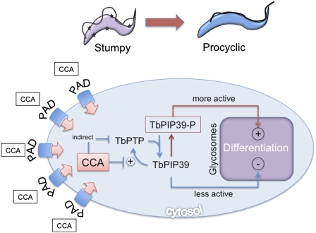
In the mammalian bloodstream, the sleeping sickness parasite Trypanosoma brucei is held poised for transmission by the activity of a tyrosine phosphatase, TbPTP1. This prevents differentiation of the transmissible "stumpy forms" until entry into the tsetse fly, whereupon TbPTP1 is inactivated and major changes in parasite physiology are initiated to allow colonization of the arthropod vector. Using a substrate-trapping approach, we identified the downstream step in this developmental signaling pathway as a DxDxT phosphatase, TbPIP39, which is activated upon tyrosine phosphorylation, and hence is negatively regulated by TbPTP1. In vitro, TbPIP39 promotes the activity of TbPTP1, thereby reinforcing its own repression, this being alleviated by the trypanosome differentiation triggers citrate and cis-aconitate, generating a potentially bistable regulatory switch. Supporting a role in signal transduction, TbPIP39 becomes rapidly tyrosine-phosphorylated during differentiation, and RNAi-mediated transcript ablation in stumpy forms inhibits parasite development. Interestingly, TbPIP39 localizes in glycosomes, peroxisome-like organelles that compartmentalize the trypanosome glycolytic reactions among other enzymatic activities. Our results invoke a phosphatase signaling cascade in which the developmental signal is trafficked to a unique metabolic organelle in the parasite: the glycosome. This is the first characterized environmental signaling pathway targeted directly to a peroxisome-like organelle in any eukaryotic cell.


Figures

Figure 1.
Figure 1.
TbPTP1 interacts with TbPIP39 in vitro and in vivo. (A) Substrate-trapping selection of TbPIP39 by TbPTP1. Wild-type (WT) or substrate-trapping recombinant TbPTP1 (D199A) was expressed as a His-tagged fusion protein and then bound to a His trap chelating column. Lysate from 5 × 10⁸ stumpy cells was then passed over the column in order to select interacting proteins. Bound material was eluted from the beads and reacted with either antibody specific for the His tag (detecting the TbPTP1 ligand), or TbPIP39. TbPIP39 was preferentially selected by the D199A TbPTP1 substrate-trapping mutant. (B) Wild-type or D199A TbPTP1-Ty was expressed in cultured bloodstream form T. brucei, which were then induced to differentiate to procyclic forms for 24 h by the addition of 6 mM cis-aconitate. Immunoprecipitation was then performed using the TY-1 epitope-specific antibody, which had been incubated either with (“blocked beads”) or without a peptide sequence recognized by the antibody. Input cell lysate, flow-through (ft), or precipitated material (beads) from each immunoprecipitation, either with or without blocking, was then reacted with the Ty1-specific antibody (to detect the ectopically expressed TbPTP1 or D199A TbPTP1) or antibodies detecting TbPIP39 or aldolase (as a negative control). The D199A-trapping mutant of TbPTP1 preferentially selected TbPIP39, whereas aldolase was not selected.
Figure 2.
Figure 2.
TbPIP39 is a phosphatase with regulatory interaction with TbPTP1. (A). Recombinant TbPIP39 was incubated with FGR kinase to generate a tyrosine-phosphorylated form over the course of 120 min. Tyrosine phosphorylation of the recombinant protein was detected using the phophotyrosine-specific antibody 4G10 (α-pTyr; Millipore). The right panel shows the same analysis using a mutant form of TbPIP39 in which the predicted tyrosine phosphorylation site Y278 is mutated to phenylalanine (F278). In this case, α-pTyr reactivity is lost. (B) Tyrosine-phosphorylated TbPIP39 is a substrate of TbPTP1. TbPIP39 was incubated with FGR kinase to generate tyrosine-phosphorylated protein, this being detected with the 4G10 antibody (α-pTyr). Upon incubation with wild-type TbPTP1, tyrosine phosphorylation of TbPIP39 is lost. In contrast, when incubated with either buffer alone or a catalytically dead (C229S) TbPTP1 mutant, TbPIP39 remains phosphorylated. (C) Activity against pNPP of recombinant TbPIP39 in the absence (column 1) or presence (column 2) of Mg2+. Column 3 shows the same assay as in column 2, using a dEdE mutant of TbPIP39. Columns 4–7 show the activity against pNPP of TbPTP1 alone (column 4) or in the presence of 1 μg (column 5), 0.1 μg (column 6), or 0.01 μg of TbPIP39. No Mg2+ was included in the reactions in columns 4–7, preventing TbPIP39 activity. Statistical analyses used a general linear model. (*) P < 0.05; (**) P < 0.001. (D) Activity against pNPP of recombinant TbPIP39 as it becomes tyrosine-phosphorylated by FGR kinase. With increasing phosphorylation, the activity increases. (Right graphs) In TbPIP39-Y278F, no enhanced activity is observed, demonstrating that tyrosine phosphorylation on Y278 is responsible for TbPIP39 regulation. The input recombinant proteins are those depicted in A. Statistical analyses used a general linear model. (*) P < 0.05.
Figure 3.
Figure 3.
TbPIP39 is a procyclic-enriched glycosomal phosphoprotein. (A) Northern blot of TbPIP39 expression in bloodstream slender (Sl), bloodstream stumpy (St), or cultured procyclic (Pro) forms. The bottom panel shows the ethidium bromide-stained rRNA, indicating loading. (B) Western blot of TbPIP39 in bloodstream slender (Sl), bloodstream stumpy (St), or cultured procyclic (Pro) forms. In stumpy forms, a lower-molecular-weight form is observed, whereas in procyclic forms, a higher-molecular-weight form predominates (arrowheads). The constitutively expressed protein TbZFP3 (Paterou et al. 2006) is included as a loading control. An empty intervening lane between the stumpy and procyclic form samples has been removed for clarity. (C) Digitonin fractionation of procyclic and stumpy form cells reacted with an antibody specific for TbPIP39, TbPIP39 (phospho-Y278), a glycosomal protein (aldolase), or a cytosolic protein (cytosolic PGK). Exposures have been adjusted to best reveal the distribution between fractions for each protein and are not equivalent between the distinct profiles. In each case, the digitonin concentration is shown with the cell extract being separated into either soluble or pelleted (organellar) fractions. TbPIP39 cofractionates with glycosomal aldolase. A quantitative analysis of the fractionation of procyclic forms is shown in Supplemental Figure 6. (D) Localization of TbPIP39 (red) with an N-terminal GFP fusion of the glycosomal protein TbPEX13 (green). The two proteins colocalize precisely. DNA of the cells was counterstained using DAPI (blue). Bar, 5 μm.
Figure 4.
Figure 4.
TbPIP39 depletion in stumpy forms inhibits differentiation. (A) Western blots of TbPIP39 from samples derived from TbPIP39-RNAi cells either induced (+Dox) or uninduced (−Dox) with doxycycline. Samples were incubated overnight at 20°C, and then proteins were isolated 0 h, 4 h, or 24 h after exposure to 0 mM, 0.1 mM, or 6 mM cis-aconitate. The bottom panels represent analysis of the same samples using an antibody against α-tubulin as a loading control. (B) Flow cytometry traces of EP procyclin expression in four independent stumpy cell populations either induced (+Dox, orange and green traces) or not (−Dox, blue and red traces) to ablate TbPIP39 by RNAi. Individual stumpy cell samples were derived from day 6 infections of mice either with or without doxycyline in their drinking water. Samples were incubated at 20°C before the addition of cis-aconitate, and flow cytometry was carried out either 4 h or 24 h later. The induced populations showed reduced differentiation at 0.1 mM (4 h and 24 h); at 6 mM, differentiation was efficient in all populations (likely enabled by the remaining ∼15% TbPIP39 in the RNAi line), although it was delayed in the induced samples. Flow cytometry traces from this experiment with cells incubated at 37°C are available in Supplemental Figure 8.
Figure 5.
Figure 5.
An intact glycosomal targeting signal is required for TbPIP39-dependent differentiation. (A) T. brucei single-marker bloodstream forms were transfected with the TbPIP39 RNAi construct used in Figure 4. The resulting transfectant cell line was then induced (blue) or not induced (red) to ablate TbPIP39 by incubation with 1 μg/mL tetracycline for 2 d. The RNAi-depleted cells and the uninduced controls were then stimulated to differentiate to procyclic forms by the addition of 6 mM cis-aconitate and the expression of the differentiation marker EP procyclin monitored by flow cytometry. Depletion of TbPIP39 resulted in reduced differentiation efficiency at 48 h after exposure to cis-aconitate. (B) The same cell line as in A was transfected with pHD451 expressing an intact recoded synthetic TbPIP39 gene, expressed under tetracycline regulation. Cells were grown with tetracycline for 2 d, allowing silencing of the endogenous TbPIP39 transcript and expression of the recoded gene. Differentiation was then monitored for EP procyclin expression 48 h after exposure to 6 mM cis-aconitate using flow cytometry. The differentiation phenotype was fully rescued. (C) As in B, but the recoded synthetic gene product lacked the ΔSRL glycosomal targeting signal and redistributed to the cytosolic fraction (Supplemental Fig. 10C). In this case, the differentiation defect was not rescued. (D) As in B, but the recoded synthetic gene product contained a Ty1 epitope tag after the ΔSRL glycosomal targeting signal. As with the truncated mutant, the protein was redistributed to the cytosolic fraction (Supplemental Fig. 10C) and the differentiation defect was not rescued. (E) As in B, but the recoded synthetic gene product was mutated for the DxDxT motif (to ExExT) to prevent catalytic activity. No rescue of the differentiation defect was observed.
Figure 6.
Figure 6.
A model depicting the regulatory interactions between TbPTP1 and TbPIP39 when exposed to the differentiation triggers citrate or cis-aconitate. The CCA signal is conveyed by PAD proteins expressed on the surface of stumpy forms. This inhibits the activation of TbPTP1 by TbPIP39, although TbPTP1 may also be inactivated indirectly. Upon the inactivation of TbPTP1, phosphorylated TbPIP39 predominates, this being generated by an unidentified protein kinase. TbPIP39, activated by phosphorylation, is directed toward the glycosomes by its C-terminal PTS1 signal, and therein promotes a differentiation response.

Similar articles

Cited by

References

    1. Albert MA, Haanstra JR, Hannaert V, Van Roy J, Opperdoes FR, Bakker BM, Michels PA 2005. Experimental and in silico analyses of glycolytic flux control in bloodstream form Trypanosoma brucei. J Biol Chem 280: 28306–28315 - PubMed
    1. Bakker BM, Mensonides FI, Teusink B, van Hoek P, Michels PA, Westerhoff HV 2000. Compartmentation protects trypanosomes from the dangerous design of glycolysis. Proc Natl Acad Sci 97: 2087–2092 - PMC - PubMed
    1. Barrett MP, Burchmore RJ, Stich A, Lazzari JO, Frasch AC, Cazzulo JJ, Krishna S 2003. The trypanosomiases. Lancet 362: 1469–1480 - PubMed
    1. Bastin P, Bagherzadeh Z, Matthews KR, Gull K 1996. A novel epitope tag system to study protein targeting and organelle biogenesis in Trypanosoma brucei. Mol Biochem Parasitol 77: 235–239 - PubMed
    1. Biebinger S, Wirtz LE, Lorenz P, Clayton C 1997. Vectors for inducible expression of toxic gene products in bloodstream and procyclic Trypanosoma brucei. Mol Biochem Parasitol 85: 99–112 - PubMed

Publication types

MeSH terms

Substances

LinkOut - more resources