. 2010 Jul 15;123(Pt 14):2369-74.
doi: 10.1242/jcs.060582. Epub 2010 Jun 15.

The nuclear protein Waharan is required for endosomal-lysosomal trafficking in Drosophila

Affiliations

The nuclear protein Waharan is required for endosomal-lysosomal trafficking in Drosophila

Mohiddin Lone et al. J Cell Sci. .

Abstract

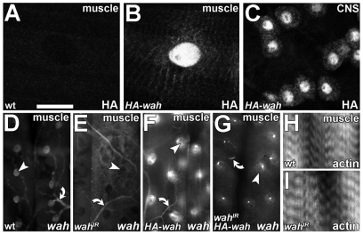
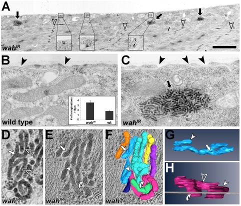
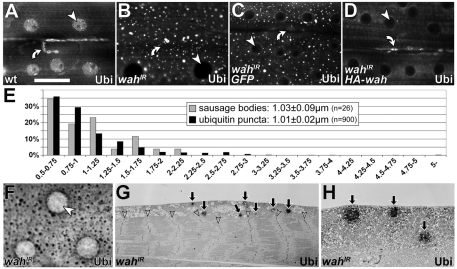
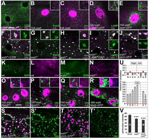
Here we report Drosophila Waharan (Wah), a 170-kD predominantly nuclear protein with two potential human homologues, as a newly identified regulator of endosomal trafficking. Wah is required for neuromuscular-junction development and muscle integrity. In muscles, knockdown of Wah caused novel accumulations of tightly packed electron-dense tubules, which we termed 'sausage bodies'. Our data suggest that sausage bodies coincide with sites at which ubiquitylated proteins and a number of endosomal and lysosomal markers co-accumulate. Furthermore, loss of Wah function generated loss of the acidic LysoTracker compartment. Together with data demonstrating that Wah acts earlier in the trafficking pathway than the Escrt-III component Drosophila Shrb (snf7 in Schizosaccharomyces pombe), our results indicate that Wah is essential for endocytic trafficking at the late endosome. Highly unexpected phenotypes result from Wah knockdown, in that the distribution of ubiquitylated cargos and endolysosomal morphologies are affected despite Wah being a predominant nuclear protein. This finding suggests the existence of a relationship between nuclear functions and endolysosomal trafficking. Future studies of Wah function will give us insights into this interesting phenomenon.


Figures

Fig. 1.
Fig. 1.
Larval expression of wah and gross muscle morphology upon Wah knockdown. (A-C) Anti-HA staining in late-larval muscles of wild type (wt; A) and in animals with HA-wah expression in muscles (B; BG57-Gal4 driver) or CNS (C; D42-Gal4 driver). (D-G) In situ hybridisation using an N-terminal wah antisense probe (see supplementary material Fig. S1B) on late-larval muscles of wild type (D), and in muscles expressing wahIR (E), HA-wah (F) or wahIR together with HA-wah (G); arrowheads indicate representative nuclei, arrows indicate potential background staining in trachea. (H,I) Wild-type (H) and wahIR-expressing (I) late-larval muscles, stained for F-actin (phalloidin). Scale bar (in A): 50 μm (for A-D,H,I); 135 μm (for D-G).
Fig. 2.
Fig. 2.
Ultrastructural analysis of wahIR-expressing muscles. (A) wahIR-expressing late-larval muscle (transverse sections) display normal contractile sarcomeres (open arrowheads point at electron-dense Z-bands); sausage bodies (black arrows and close-ups) accumulate predominantly in the sarcoplasm. (B,C) Tubular surface invaginations (black arrowheads) are less abundant in wild-type (wt) than in wahIR-expressing muscles (inset in B). Black arrow in C indicates a sausage body. (D) Higher-resolution view of a sausage body showing branch points (white arrow) and even spacing between electron tubules (double chevrons). (E-H) EM tomogram of a sausage body (E) and its 3D reconstruction with individual tubules shown in different colours (F-H); see supplementary material Movies 1 and 2); tubules penetrating the image plane (grey line in H) perpendicularly (curved arrow in H) appear as pseudo-vesicular structures in horizontal view (curved arrow in E). Symbols are used consistently throughout the figure. Scale bar (in A): 3 μm (for A); 0.6 μm (for B,C); 120 nm (for D-F); 100 nm (for G); 50 nm (for H).
Fig. 3.
Fig. 3.
Characterisation of wahIR-induced ubiquitin puncta. (A-D) Anti-ubiquitin staining in the sarcoplasm of late-larval muscles in wild type (wt; A), and in muscles expressing wahIR (B), coexpressing wahIR with a GFP construct (C) or coexpressing wahIR with HA-wah (D); arrowheads point at nuclei, curved arrows at neuromuscular junctions (both of which lack ubiquitin upon muscular knockdown of Wah). (E) Distributions of diameters (larger than 0.5 μm) of sausage bodies and ubiquitin puncta; inset indicates the average diameters. (F-H) Pre-embedding immuno-EM for ubiquitin in wahIR-expressing late-larval muscles (diaminobenzidine staining catalysed by horseradish peroxidase); resin-embedded specimens show the same staining pattern as fluorescently labelled muscles (compare F and B); ubiquitin-positive patches localise predominantly in the sarcoplasm (G,H), as is similarly found for sausage bodies (compare Fig. 2A). Arrows indicate sausage bodies; arrowheads indicate nuclei. Scale bar (in A): 50 μm (for A-D,F); 12 μm (for G); 2.2 μm (for H).
Fig. 4.
Fig. 4.
Refining insights into endosomal functions of Wah. All images show horizontal views of late-larval muscles at the level of the sarcoplasm; genotypes or expressed constructs are indicated bottom left, detected labels bottom right. (A-J) Localisation of the membrane marker mCD8-GFP (CD8) and the endosomal and/or lysosomal markers Rab5, Hrs, LAMP-GFP (LAMP) and Spinster-GFP (Spin) in control animals (A-E), and upon knockdown of Wah (F-J); none of the endosomal markers colocalised with HA-Wah (B-E); upon knockdown of Wah, CD8-GFP and endosomal markers accumulate and closely correlate with ubiquitin puncta (F-J). (K-N) Puncta of LysoTracker staining detected by live imaging of wild-type muscles (lyso; K) is abolished upon wahIR expression (M); L and N show specimens from the same batch subsequently stained for ubiquitin. (O-R) Targeted expression of constructs functionally interfering with endosomal trafficking: dominant-negative and activated Rab5 (Rab5SN and Rab5QL, respectively), activated Rab7 (Rab7QL), and dominant-negative snf7 (shrb-GFP); these conditions cause accumulations of ubiquitin (O,P,Q,S), but do not affect the localisation of cytoplasmic HA-Wah puncta (O-R). (S,T) Shrb-GFP co-clusters with ubiquitin around nuclei (S,S′); coexpression of wahIR suppresses perinuclear build-up of ubiquitin (T,T′; see also V) and its localisation to the Shrb-GFP compartment (see also U). (U) Pearson's colocalisation correlation coefficients (1=perfect colocalisation, 0=random distribution, −1=perfect exclusion) for all double-stained images of this plate, as indicated by letters below bars; HA-Wah fails to colocalise with endosomal markers (upper graph, left) and with ubiquitin accumulations induced by endosomal manipulations (upper graph, right); ubiquitin puncta induced by wahIR (lower graph, left) or Shrb-GFP (lower graph, right) colocalise with endosomal markers. ***P≤0.001, Mann-Whitney Rank Sum Test. (V) Pixel intensities of ubiquitin staining measured in circular ROIs of 35-μm diameter concentrically around nuclei; bars represent the means of respective histograms. ***P≤0.001, Mann-Whitney Rank Sum Test. Scale bar (in A): 30 μm (for K-N); 15 μm (for all other images; insets are 200% enlarged).

References

    1. Arents G., Moudrianakis E. N. (1995). The histone fold: a ubiquitous architectural motif utilized in DNA compaction and protein dimerization. Proc. Natl. Acad. Sci. USA 92, 11170-11174 - PMC - PubMed
    1. Askree S. H., Yehuda T., Smolikov S., Gurevich R., Hawk J., Coker C., Krauskopf A., Kupiec M., McEachern M. J. (2004). A genome-wide screen for Saccharomyces cerevisiae deletion mutants that affect telomere length. Proc. Natl. Acad. Sci. USA 101, 8658-8663 - PMC - PubMed
    1. Bate M. (1993). The mesoderm and its derivatives. In The development of Drosophila melanogaster, vol. 2 (eds Bate M., Martínez Arias A.), pp. 1013-1090 Cold Spring Harbor: CSH Laboratory Press;
    1. Baudhuin P., Beaufay H., De Duve C. (1965). Combined biochemical and morphological study of particulate fractions from rat liver. Analysis of preparations enriched in lysosomes or in particles containing urate oxidase, D-amino acid oxidase, and catalase. J. Cell Biol. 26, 219-243 - PMC - PubMed
    1. Berndsen C. E., Tsubota T., Lindner S. E., Lee S., Holton J. M., Kaufman P. D., Keck J. L., Denu J. M. (2008). Molecular functions of the histone acetyltransferase chaperone complex Rtt109-Vps75. Nat. Struct. Mol. Biol. 15, 948-956 - PMC - PubMed

Publication types

MeSH terms

Substances