

Anatomical variations of hepatic arterial system, coeliac trunk and renal arteries: an analysis with multidetector CT angiography
- PMID: 20551256
- PMCID: PMC3473504
- DOI: 10.1259/bjr/21236482
Anatomical variations of hepatic arterial system, coeliac trunk and renal arteries: an analysis with multidetector CT angiography
Abstract
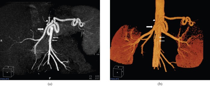
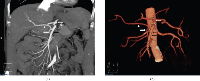
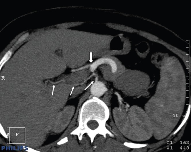
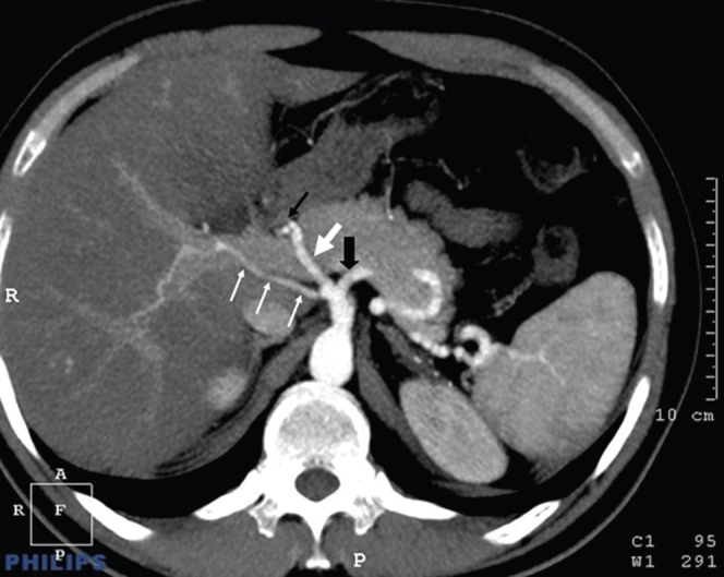
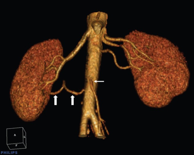
The purpose of our investigation was to determine the anatomical variations in the coeliac trunk-hepatic arterial system and the renal arteries in patients who underwent multidetector CT (MDCT) angiography of the abdominal aorta for various reasons. A total of 100 patients were analysed retrospectively. The coeliac trunk, hepatic arterial system and renal arteries were analysed individually and anatomical variations were recorded. Statistical analysis of the relationship between hepatocoeliac variations and renal artery variations was performed using a chi(2) test. There was a coeliac trunk trifurcation in 89% and bifurcation in 8% of the cases. Coeliac trunk was absent in 1%, a hepatosplenomesenteric trunk was seen in 1% and a splenomesenteric trunk was present in 1%. Hepatic artery variation was present in 48% of patients. Coeliac trunk and/or hepatic arterial variation was present in 23 (39.7%) of the 58 patients with normal renal arteries, and in 27 (64.3%) of the 42 patients with accessory renal arteries. There was a statistically significant correlation between renal artery variations and coeliac trunk-hepatic arterial system variations (p = 0.015). MDCT angiography permits a correct and detailed evaluation of hepatic and renal vascular anatomy. The prevalence of variations in the coeliac trunk and/or hepatic arteries is increased in people with accessory renal arteries. For that reason, when undertaking angiographic examinations directed towards any single organ, the possibility of variations in the vascular structure of other organs should be kept in mind.
Figures
References
-
- Munshi IA, Fusco D, Tashjian D, Kirkwood JR, Polga J, Wait RB. Occlusion of an aberrant right hepatic artery, originating from the superior mesenteric artery, secondary to blunt trauma. J Trauma 2000;48:325–6 - PubMed
-
- Rela M, McCall JL, Karani J, Heaton ND. Accessory right hepatic artery arising from the left. Transplantation 1998;66:792–4 - PubMed
-
- Fox M, Yalin R. Renal transplantation with multiple arteries. Br J Urol 1979;51:333–6 - PubMed
-
- Sampaio FJB, Passos MARF. Renal arteries: anatomic study for surgical and radiological practice. Surg Radiol Anat 1992;14:113–17 - PubMed
-
- Uflacker R. Atlas of vascular anatomy: an angiographic approach. Baltimore: Williams & Wilkins, 1997
