. 2010 Sep 1;334(3):761-74.
doi: 10.1124/jpet.110.168211. Epub 2010 Jun 15.

Negative allosteric modulators that target human alpha4beta2 neuronal nicotinic receptors

Affiliations

Negative allosteric modulators that target human alpha4beta2 neuronal nicotinic receptors

Brandon J Henderson et al. J Pharmacol Exp Ther. .

Abstract

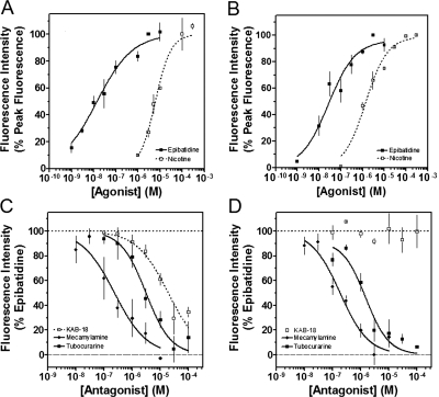
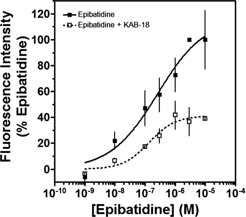
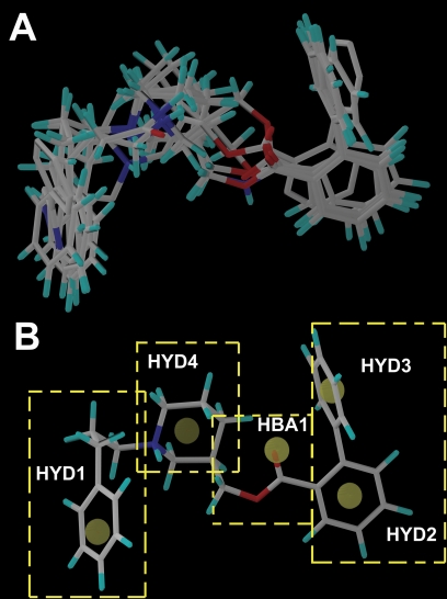
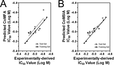
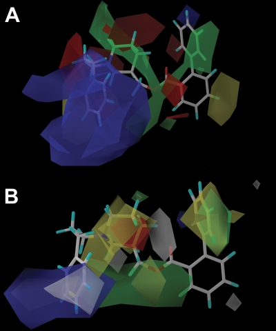
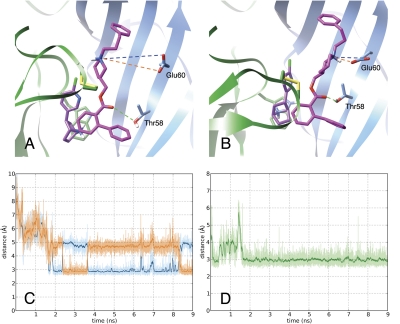
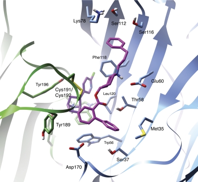
Allosteric modulation of neuronal nicotinic acetylcholine receptors (nAChRs) is considered to be one of the most promising approaches for therapeutics. We have previously reported on the pharmacological activity of several compounds that act as negative allosteric modulators (NAMs) of nAChRs. In the following studies, the effects of 30 NAMs from our small chemical library on both human alpha4beta2 (Halpha4beta2) and human alpha3beta4 (Halpha3beta4) nAChRs expressed in human embryonic kidney ts201 cells were investigated. During calcium accumulation assays, these NAMs inhibited nAChR activation with IC(50) values ranging from 2.4 microM to more than 100 microM. Several NAMs showed relative selectivity for Halpha4beta2 nAChRs with IC(50) values in the low micromolar range. A lead molecule, KAB-18, was identified that shows relative selectivity for Halpha4beta2 nAChRs. This molecule contains three phenyl rings, one piperidine ring, and one ester bond linkage. Structure-activity relationship (SAR) analyses of our data revealed three regions of KAB-18 that contribute to its relative selectivity. Predictive three-dimensional quantitative SAR (comparative molecular field analysis and comparative molecular similarity indices analysis) models were generated from these data, and a pharmacophore model was constructed to determine the chemical features that are important for biological activity. Using docking approaches and molecular dynamics on a Halpha4beta2 nAChR homology model, a binding mode for KAB-18 at the alpha/beta subunit interface that corresponds to the predicted pharmacophore is described. This binding mode was supported by mutagenesis studies. In summary, these studies highlight the importance of SAR, computational, and molecular biology approaches for the design and synthesis of potent and selective antagonists targeting specific nAChR subtypes.


Figures

Fig. 1.
Fig. 1.
Concentration–response effects of agonists, antagonists, and KAB-18 on Hα4β2 and Hα3β4 nAChRs. A and B, the concentration–response effects of nicotine (□) and epibatidine (■) were investigated by using HEK ts201 cells expressing Hα4β2 and Hα3β4 nAChRs. Data are expressed as a percentage of peak fluorescence responses for either 3 μM epibatidine (epibatidine curves) or 100 μM nicotine (nicotine curves). C and D, tubocurarine (■), mecamylamine (♦), and KAB-18 (□) were investigated by using HEK ts201 cells expressing Hα4β2 and Hα3β4 nAChRs. Epibatidine (3 μM) was used as the agonist, and data are expressed as a percentage of peak fluorescence responses for 3 μM epibatidine. The dotted and dashed horizontal lines show the 100% control response and 100% inhibition, respectively. Values represent means ± S.E.M.s (n = 4–10).
Fig. 2.
Fig. 2.
Concentration–response effects of epibatidine in the absence or presence of KAB-18. The concentration-response effects of epibatidine were investigated in the absence (■) or presence (□) of 30 μM KAB-18 by using HEK ts201 cells expressing Hα4β2 nAChRs. Values represent means ± SEMs (n = 4).
Fig. 3.
Fig. 3.
Alignment of the molecules and the proposed pharmacophore targeting the negative allosteric site of the Hα4β2 nAChR. A, the alignment of the NAMs used in model generation is shown in capped stick representation. B, the features of the pharmacophore model generated using GASP are illustrated using KAB-18. Four hydrophobic features (HYD1, HYD2, HYD3, and HYD4) and a hydrogen bond acceptor (HBA1) feature of the pharmacophore are marked. The yellow dashed boxes outline the four regions used in the SAR studies.
Fig. 4.
Fig. 4.
Predicted and experimentally derived functional IC50 values of training set and test set compounds using CoMFA model (A) or CoMSIA model (B) were compared and linear regression analysis was performed. Log IC50 values of training set compounds are listed in Table 6, and log IC50 values of test set compounds are listed in Table 7.
Fig. 5.
Fig. 5.
CoMFA and CoMSIA models of NAM binding. A, CoMFA coefficient contours map aligned with KAB-18. The contours of the steric map are shown in yellow and green, and the contours of the electrostatic map are shown in red and blue. Greater potency (lower IC50 values) is correlated with less bulk near yellow, more bulk near green, more negative charge near red, and more positive charge near blue. B, CoMSIA contours aligned with KAB-18. The contours of the hydrophobic map are shown in white, yellow, and green, and the contours of the HBA map are shown in red and blue. Greater potency (lower IC50 values) is correlated with less bulk near yellow, more bulk near green and white, more HBA near blue, and less HBA near red.
Fig. 6.
Fig. 6.
Stability of KAB-18 at its proposed binding site. A and B, MD simulation of the Hα4β2 model was used to demonstrate initial docking mode (A) and induced binding mode at 7 ns (B) of KAB-18 (magenta) to the epibatidine-bound (purple) interface of the Hα4 (green ribbon) and Hβ2 (blue ribbon) subunits. Dotted lines identify key polar interactions between the ligand and the receptor. C and D, interatomic distances over time between the nitrogen of the positively charged piperidine and the two oxygen atoms of β2Glu60 (orange and blue labeling) and the keto group of the ester linkage and the hydroxyl-oxygen of β2Thr58 (green labeling), respectively.
Fig. 7.
Fig. 7.
Identification of ligand–receptor contacts of KAB-18 in the Hα4β2 nAChR model. A snapshot at 7.0 ns from MD simulation shows the induced binding mode of KAB-18 (magenta) to the epibatidine-bound (purple) interface of the Hα4 (green ribbon) and Hβ2 (blue ribbon) subunits. Key amino acids contributing to the binding of KAB-18 are shown as stick figures.

Similar articles

Cited by

References

    1. Anand R, Conroy WG, Schoepfer R, Whiting P, Lindstrom J. (1991) Neuronal nicotinic acetylcholine receptors expressed in Xenopus oocytes have a pentameric quaternary structure. J Biol Chem 266:11192–11198 - PubMed
    1. Arias HR. (1998) Binding sites for exogenous and endogenous non-competitive inhibitors of the nicotinic acetylcholine receptor. Biochim Biophys Acta 1376:173–220 - PubMed
    1. Bergmeier SC, Ismail KA, Arason KM, McKay S, Bryant DL, McKay DB. (2004) Structure activity studies of ring E analogues of methyllycaconitine. Part 2: synthesis of antagonists to the α3β4 nicotinic acetylcholine receptors through modifications to the ester. Bioorg Med Chem Lett 14:3739–3742 - PubMed
    1. Bergmeier SC, Lapinsky DJ, Free RB, McKay DB. (1999) Ring E analogs of methyllycaconitine (MLA) as novel nicotinic antagonists. Bioorg Med Chem Lett 9:2263–2266 - PubMed
    1. Bryant DL, Free RB, Thomasy SM, Lapinsky DJ, Ismail KA, McKay SB, Bergmeier SC, McKay DB. (2002) Structure-activity studies with ring E analogues of methyllycaconitine on bovine adrenal α3β4* nicotinic receptors. Neurosci Res 42:57–63 - PubMed

Publication types

MeSH terms