. 2010 Sep 3;285(36):28141-55.
doi: 10.1074/jbc.M109.043679. Epub 2010 Jun 15.

Knockdown of zebrafish lumican gene (zlum) causes scleral thinning and increased size of scleral coats

Affiliations

Knockdown of zebrafish lumican gene (zlum) causes scleral thinning and increased size of scleral coats

Lung-Kun Yeh et al. J Biol Chem. .

Abstract

The lumican gene (lum), which encodes one of the major keratan sulfate proteoglycans (KSPGs) in the vertebrate cornea and sclera, has been linked to axial myopia in humans. In this study, we chose zebrafish (Danio rerio) as an animal model to elucidate the role of lumican in the development of axial myopia. The zebrafish lumican gene (zlum) spans approximately 4.6 kb of the zebrafish genome. Like human (hLUM) and mouse (mlum), zlum consists of three exons, two introns, and a TATA box-less promoter at the 5'-flanking region of the transcription initiation site. Sequence analysis of the cDNA predicts that zLum encodes 344 amino acids. zLum shares 51% amino acid sequence identity with human lumican. Similar to hLUM and mlum, zlum mRNA is expressed in the eye and many other tissues, such as brain, muscle, and liver as well. Transgenic zebrafish harboring an enhanced GFP reporter gene construct downstream of a 1.7-kb zlum 5'-flanking region displayed enhanced GFP expression in the cornea and sclera, as well as throughout the body. Down-regulation of zlum expression by antisense zlum morpholinos manifested ocular enlargement resembling axial myopia due to disruption of the collagen fibril arrangement in the sclera and resulted in scleral thinning. Administration of muscarinic receptor antagonists, e.g. atropine and pirenzepine, effectively subdued the ocular enlargement caused by morpholinos in in vivo zebrafish larvae assays. The observation suggests that zebrafish can be used as an in vivo model for screening compounds in treating myopia.


Figures

FIGURE 1.
FIGURE 1.
Schematic diagram showing the organization of the zlum gene. The zebrafish lumican gene is 11 kb upstream from the keratocan gene. It contains three exons and two introns. The figure shows the zebrafish lumican gene drawn to scale. Blank boxes indicate the coding region of the mRNA. The translation start and stop codons are indicated by ATG and TAA, respectively.
FIGURE 2.
FIGURE 2.
Neighbor-joining phylogenic tree of the lumican protein. Phylogenetic tree of lumican protein from 18 different species. The numbers at the nodes represent the statistical confidence estimates computed by the bootstrap procedure. Bootstrap values were calculated from 100 replicates, and values of >50% are indicated at each divergence point. The following are cited: Canis familiaris (dog); Equus caballus (horse); Sus scrofa (pig); Bos taurus (cow); Mus musculus (mouse); Rattus norvegicus (rat); Pan troglodytes (chimpanzee); Macaca fascicularis (macaque); Macaca mulatta (rhesus mulatta); Homo sapiens (human); Pongo abelii (Sumatran orangutan); Ornithorhynchus anatinus (platypus); Salmo salar (Atlantic salmon); Danio rerio (zebrafish); Silurana tropicalis (Xenopus); Taeniopygia guttata (zebra finch); Coturnix japonica (Japanese quail); Gallus gallus (chicken). The tree was constructed with Geneious Pro software (version 4.7).
FIGURE 3.
FIGURE 3.
zlum expression by RT-PCR analyses. RT-PCR was carried out using total RNAs from adult fish eye (lane 1), brain (lane 2), heart (lane 3), liver (lane 4), gut (lane 5), muscle (lane 6), and fin (lane 7). Note that amplified zlum PCR product could be found not only in the eye but also in other tissues (bottom panel).
FIGURE 4.
FIGURE 4.
In situ hybridization analyses of zLum mRNA in early zebrafish larvae (whole mount) and adult fish cornea (sections). Hybridization signals were demonstrated with anti-digoxigenin antibody-alkaline phosphatase conjugates. A, zLum mRNA was detected in zebrafish larvae (24 hpf). B, in older zebrafish larvae (48 hpf), the zLum mRNA was detected in the eyes and all the major subdivisions of the embryonic central nervous system, including the fore-, mid-, and hindbrain and the spinal cord. C, strong hybridization signals could be also found in corneal tissue at 48 hpf. D, no hybridization signals could be detected in sense probe hybridization group at the 48-hpf larval stage. E, in the zebrafish eye, zLum mRNA was predominantly detected in the corneal stromal layer. F, higher magnification showed that hybridization-positive signals are observed in the corneal stromal layer. G, zLum mRNA was also detected in the periocular extracellular matrix. H, higher magnification showed that hybridization-positive signals are observed in the scleral layer (arrow). I–L, no hybridization signals could be detected in the adult eye (I), cornea (J), or sclera (K and L) using a sense probe in hybridization. Sections were counterstained with fast red. (Scale bars, 100 μm, E, G, I, and K; and 50 μm, F, H, J, and L.)
FIGURE 5.
FIGURE 5.
Immunohistochemistry staining pattern of zebrafish eye using the epitope-specific anti-zLum antibody. A, lumican protein was found in zebrafish eye tissue. B, note that lumican protein was found mainly in the corneal stromal layer. C and D, lumican protein was also found in the scleral tissue (arrow). E–H, no immunoreactivity was detected in the negative control group. (E, whole eye; F, corneal tissue; G and H, scleral tissue.) I–P, tissue sections treated with (M–P) and without (I–L) keratanase and stained with an anti-keratan sulfate antibody are shown. I and J, tissue sections showed that keratan sulfate chains existed mainly in the corneal stromal layer. K and L, keratan sulfate chains did not exist in the scleral layer. M–P, there was nearly no immunoreactive reaction in tissue sections treated with keratanase and stained with anti-keratan sulfate antibody. (Scale bars, 200 μm, A, E, I, and M; scale bars, 100 μm, C, G, K, and O; scale bars, 50 μm, B, D, F, H, J, L, N, and P; and arrow indicates scleral tissue ins D, H, L, and P.)
FIGURE 6.
FIGURE 6.
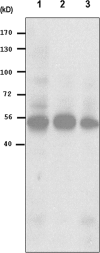
Western blot analysis of zebrafish lumican. Total proteins extracted from adult zebrafish eyes by lysis buffer were subjected to 10% SDS-PAGE followed by Western blotting. Samples without treatment (lane 1) and with either keratanase (lane 2) or endo-β-galactosidase treatment (lane 3) were probed with a rabbit anti-zebrafish lumican antibody. The result showed multiple bands with a similar smearing pattern without enzyme digestion (lane 1), whereas one ∼50-kDa band was visualized after treatment with keratanase (lane 2) and endo-β-galactosidase (lane 3). These data demonstrate that the zebrafish lumican protein contains keratan sulfate chains.
FIGURE 7.
FIGURE 7.
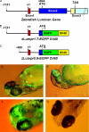
Generation of transgenic fish harboring zlumpr1.7-EGFP SV40 and zlumpr0.5-EGFPSV40. A, schematic representation of the zebrafish lumican gene. B, structure of zlumpr1.7-EGFP SV40 (3.3 kb). It contains a 1.7-kb 5′-regulatory region of the zlum gene, the untranslated region of exon 1 (844 bp), an SV40 polyadenylation signal, and the pEGFP vector sequence. C, structure of zlumpr0.5-EGFPSV40 (2.0 kb). It contains a 0.5-kb 5′-regulatory region of the zlum gene, the untranslated region of exon 1 (844 bp), an SV40 polyadenylation signal, and the pEGFP vector sequence. D, EGFP expression was observed in the 3 dpf zebrafish after injecting linearized zlumpr1.7-EGFP SV40 DNA fragment. E, under different observation view of the same fish (3 dpf), strong EGFP expression was detected in corneal tissue. F, EGFP was expressed at the 7 dpf stage. G, as a control, no EGFP was detected after injecting the linearized zlumpr0.5-EGFP SV40 DNA fragment.
FIGURE 8.
FIGURE 8.
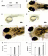
Ocular enlargement developed because of the reduction of zLum protein by morpholino microinjection. A, Western blot was carried out using embryo lysates from wild type embryos (1st lane) and zlum-MO-injected 3-dpf embryos (2nd lane). It showed that zLum protein decreased after zlum-MO was injected. B, this figure showed the definition of axial length (AL) and diameter (D) of eye in zebrafish. C, RS-MO-injected embryos showed normal phenotypes as a control group at 22 hpf stages. D, developmental delay was found in the zlum-MO-injected group at 22 hpf stages. E, normal phenotype was noted in the RS-MO-injected group at the 7 dpf stage. F, significant ocular enlargement was noted in the zlum-MO-injected group at the 7 dpf stage. G, significantly increased axial length was noted in the zlum-MO-injected group as compared with the RS-MO-injected embryos and wild type embryos at 7 dpf (respectively, p < 0.05). H, significantly increased eyeball diameter was also noted in the zlum-MO-injected group compared with the RS-MO-injected embryos and wild type embryos at 7 dpf (respectively, p < 0.05).
FIGURE 9.
FIGURE 9.
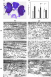
zlum-MO knockdown induces ultrastructural changes in the CS, AS, and PS. A, WT fish at 12 dpf stage in toluidine blue staining. The figure indicates CS, AS, and PS. B, diameters of collagen fibril were analyzed in the corneal stroma, anterior and posterior sclera of 12 dpf-old-wild type, and zlum-MO-injected group. Significant increases in collagen fibril diameter of corneal stroma and anterior sclera are noted in the zlum-MO group, and the diameter of collagen fibril in the posterior sclera is not significantly different in both groups. C–H, morphological comparison of collagen fibril architecture in the corneal stroma (C and D), anterior scleral tissue (E and F), and posterior scleral tissue (G and H) between the control group (C, E, and G) and zlum-MO-injected group (D, F, and H) at the 12 dpf stage. C, TEM micrograph showing regular and smaller fibril architecture of collagen localized in the corneal stroma of the wild type group. D, irregular arrangement and increasing collagen fibril diameter was found in the corneal stroma of the zlum-MO-injected group. E, TEM micrograph showing relatively regular fibril architecture of collagen localized in the anterior sclera of the wild type group. F, irregular collagen fibrils with increasing fibril diameter were noted in the anterior sclera of the zlum-MO-injected group. G, top is adjacent to the retina. TEM micrograph showing fibril architecture of collagen localized in the posterior sclera of the wild type group. H, top is adjacent to the retina. TEM micrograph showing irregular and little collagen fibril architecture was noted in the posterior sclera of the zlum-MO-injected group. (scale bar, C–H, 100 nm.)
FIGURE 10.
FIGURE 10.
Ultrastructure changes in scleral thinning in zlum-MO group. A, top is adjacent to the retina. Two to three layers of scleral fibroblastic cells with collagen fibril formation between the layers was found at the posterior sclera of the WT fish at 7 dpf stage. B, top is adjacent to the retina. Only one to two layers of fibroblastic cells at the posterior sclera of the zlum-MO-injected fish at 7 dpf stage is shown. C, scleral thinning was observed obviously in the zlum-MO-injected fish at 7 dpf stage. The phenomenon was much more prominent in the zlum-MO-injected fish at 12 dpf stage. In particular, significant scleral thinning was observed in the posterior sclera of the zlum-MO-injected fish at 7 and 12 dpf stage as compared with wild type group. (Scale bar, A and B, 1.5 μm.)
FIGURE 11.
FIGURE 11.
Zebrafish larvae assay for drug screen. A, normal phenotype of WT fish at 7 dpf stage. B, normal phenotype of RS-MO-injected embryos at 7 dpf stage. C, significantly enlarged eyeball of zlum-MO-injected fish at 7 dpf stage. D, significant decrease in ocular enlargement was noted in the zlum-MO-injected larvae at 7 dpf stage after treating with 0.5% atropine for 2 days. E, decrease in ocular enlargement was also found in the zlum-MO-injected larvae at 7 dpf stage after treating with 0.25% pirenzepine (P) for 2 days. F, no obvious changes in the phenotypes of zlum-MO-injected fish at 7 dpf stage after treating with 0.01% methoctramine (M).
FIGURE 12.
FIGURE 12.
Zebrafish drug screen assay. A and B show the definition of outer margin of RPE (red color) and diameter (D) of scleral coat (green color) in zebrafish. C, significant decrease in excessive axial elongation in the zlum-MO-injected fish at the 7 dpf stage after treating with 0.5% atropine (A) and 0.25% pirenzepine (P), whereas no obvious changes in excessive axial elongation after treating with 0.01% methoctramine (M). 1st lane, WT; 2nd lane, MO + 0.5% atropine; 3rd lane, MO + 0.25% pirenzepine; 4th lane, MO + 0.01% methoctramine; 5th lane, MO. Significant decrease in the diameter of scleral coat of the zlum-MO-injected fish at the 7 dpf stage after treating with 0.5% atropine and 0.25% pirenzepine, whereas no obvious changes in the zlum-MO-injected group treated with 0.01% methoctramine. 6th lane, WT; 7th lane, MO + 0.5% atropine; 8th lane, MO + 0.25% pirenzepine; 9th lane, MO + 0.01% methoctramine; 10th lane, MO. D, significant decrease in the ratio of RPE/scleral coat (%) was noted during ocular enlargement developed due to the reduction of zLum protein. Some muscarinic receptor antagonists (atropine and pirenzepine) attenuate the decreasing ratio of RPE/scleral coat due to the reduction of zLum protein, whereas there was no obvious changes in the decreased ratio of RPE/scleral coat in the methoctramine-treated group. 1st lane, WT; 2nd lane, MO + 0.5% atropine; 3rd lane, MO + 0.25% pirenzepine; 4th lane, MO + 0.01% methoctramine; 5th lane, MO.

Similar articles

Cited by

References

    1. Lin L. L., Shih Y. F., Hsiao C. K., Chen C. J., Lee L. A., Hung P. T. (2001) J. Formos. Med. Assoc. 100, 684–691 - PubMed
    1. Kempen J. H., Mitchell P., Lee K. E., Tielsch J. M., Broman A. T., Taylor H. R., Ikram M. K., Congdon N. G., O'Colmain B. J. (2004) Arch. Ophthalmol. 122, 495–505 - PubMed
    1. McBrien N. A., Metlapally R., Jobling A. I., Gentle A. (2006) Invest. Ophthalmol. Vis. Sci. 47, 4674–4682 - PubMed
    1. Deere M., Johnson J., Garza S., Harrison W. R., Yoon S. J., Elder F. F., Kucherlapati R., Hook M., Hecht J. T. (1996) Genomics 38, 399–404 - PubMed
    1. Fisher L. W., Heegaard A. M., Vetter U., Vogel W., Just W., Termine J. D., Young M. F. (1991) J. Biol. Chem. 266, 14371–14377 - PubMed

Publication types

MeSH terms

Associated data