. 2010 Aug 27;285(35):27289-27301.
doi: 10.1074/jbc.M110.131003. Epub 2010 Jun 15.

Postendocytic sorting of constitutively internalized dopamine transporter in cell lines and dopaminergic neurons

Affiliations

Postendocytic sorting of constitutively internalized dopamine transporter in cell lines and dopaminergic neurons

Jacob Eriksen et al. J Biol Chem. .

Abstract

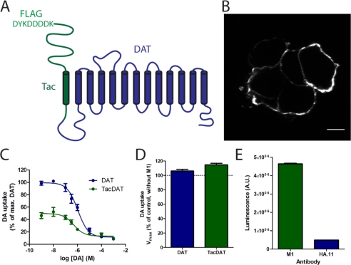
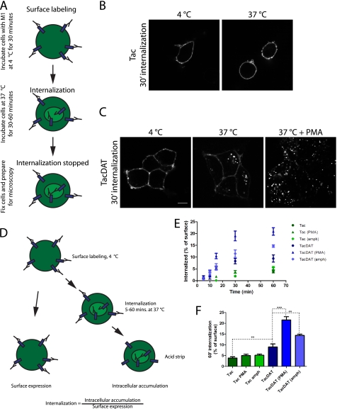
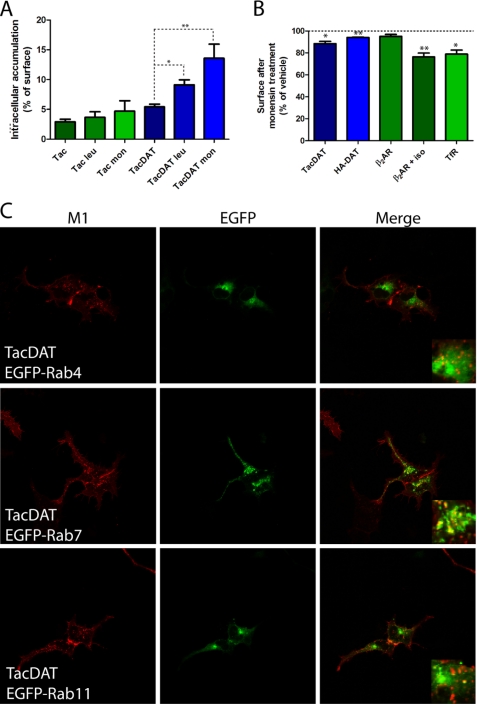
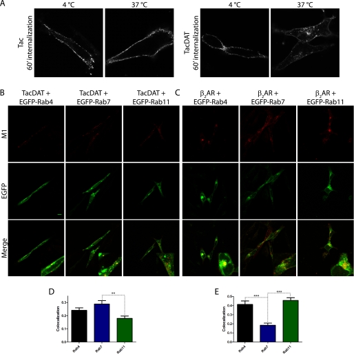
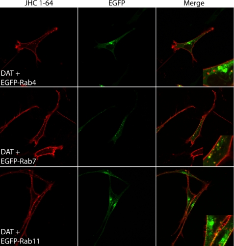
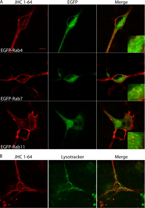
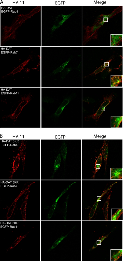
The dopamine transporter (DAT) mediates reuptake of released dopamine and is the target for psychostimulants, such as cocaine and amphetamine. DAT undergoes marked constitutive endocytosis, but little is known about the fate and sorting of the endocytosed transporter. To study DAT sorting in cells lines, we fused the one-transmembrane segment protein Tac to DAT, thereby generating a transporter (TacDAT) with an extracellular antibody epitope suited for trafficking studies. TacDAT was functional and endocytosed constitutively in HEK293 cells. According to an ELISA-based assay, TacDAT intracellular accumulation was increased by the lysosomal protease inhibitor leupeptin and by monensin, an inhibitor of lysosomal degradation and recycling. Monensin also reduced TacDAT surface expression consistent with partial recycling. In both HEK293 cells and in the dopaminergic cell line 1Rb3An27, constitutively internalized TacDAT displayed primary co-localization with the late endosomal marker Rab7, less co-localization with the "short loop" recycling marker Rab4, and little co-localization with the marker of "long loop" recycling endosomes, Rab11. Removal by mutation of N-terminal ubiquitination sites did not affect this sorting pattern. The sorting pattern was distinct from a bona fide recycling membrane protein, the beta(2)-adrenergic receptor, that co-localized primarily with Rab11 and Rab4. Constitutively internalized wild type DAT probed with the fluorescently tagged cocaine analogue JHC 1-64, exhibited the same co-localization pattern as TacDAT in 1Rb3An27 cells and in cultured midbrain dopaminergic neurons. We conclude that DAT is constitutively internalized and sorted in a ubiquitination-independent manner to late endosomes/lysosomes and in part to a Rab4 positive short loop recycling pathway.


Figures

FIGURE 1.
FIGURE 1.
Thirteen-transmembrane segment fusion protein between DAT and Tac contains high affinity FLAG epitope and is functionally expressed in plasma membrane of HEK293 cells. A, TacDAT is a head-to-tail fusion of hDAT (blue) and Tac (green) with an N-terminal M1 antibody FLAG epitope, DYKDDDDK. B, non-permeabilizing immunostaining of HEK293 cells transfected with TacDAT using an M1 antibody recognizing the FLAG epitope. Scale bar, 10 μm. C, [3H]dopamine (DA) uptake experiments in HEK293 cells transfected with TacDAT (green) or hDAT (blue). The data are shown as relative uptake in percentage of [3H]dopamine uptake in wild type DAT (means ± S.E., n = 3 of triplicate determinations). D, [3H]dopamine uptake after preincubating HEK293 cells expressing TacDAT or hDAT for 30 min at 4 °C with M1 antibody. Data are means ± S.E. of n = 3. E, surface HA.11 and M1 ELISA signals from HEK293 cells expressing TacDAT with an inserted HA tag in the second extracellular loop (Tac-HA-DAT). Values are means ± S.E. of n = 3. A.U., arbitrary units.
FIGURE 2.
FIGURE 2.
Visualizing and quantifying TacDAT internalization using M1 antibody-based internalization assays. A, to detect TacDAT internalization, we established a conventional antibody internalization assay in which we first labeled the cells with M1 antibody at 4 °C to label surface TacDAT at a temperature that blocked trafficking. Next, the cells were incubated at 37 °C in new medium for 30–60 min to allow internalization of TacDAT. For surface detection alone, the cells were kept at 4 °C. Subsequently, cells were fixed, permeabilized, and incubated with secondary fluorophore-conjugated antibody. The fluorescence was visualized using confocal microscopy. B, confocal images of HEK293 cells expressing Tac assayed as just described. C, the same assay with HEK293 cells expressing TacDAT. During the 30-min incubation at 37 °C, cells were incubated with or without the PKC activator PMA (1 μm). The images are representative of at least three similar experiments. D, brief description of ELISA-based internalization assay. As for the conventional microscopy-based internalization assay described above, cells were first labeled with M1 at 4 °C and then either incubated at 4 °C for surface detection or incubated at 37 °C for various periods to allow internalization. After the internalization period, the cells were placed on ice, and the surface antibody was stripped off using an acid strip buffer. After fixation, the cells were permeabilized, and the intracellular accumulated antibody was detected with a horseradish peroxidase-conjugated secondary antibody. The internalization signal was expressed as the proportion of the start surface signal. E, intracellular accumulation assessed by ELISA (as just described) in HEK293 cells transiently expressing TacDAT or Tac. Internalization was performed without or with PMA (1 μm) or amphetamine (10 μm) for the indicated times. Values are means ± S.E. of n = 6. F, intracellular accumulation after 60 min of internalization (means ± S.E. of n = 6; **, p < 0.01; ***, p < 0.001; one-way ANOVA with Bonferroni's multiple comparison test). amph, amphetamine; ′, minutes.
FIGURE 3.
FIGURE 3.
Monensin and leupeptin affect DAT trafficking, and TacDAT co-localizes with late endosomal marker Rab7 in HEK293 cells. A, intracellular accumulation measured by ELISA (described in Fig. 2D) in HEK293 cells transfected with Tac or TacDAT. The protease inhibitor leupeptin (leu) (100 μg/ml) or the recycling inhibitor monensin (mon) (25 μm) was included as indicated during the 1 h of internalization (means ± S.E. of n = 4; *, p < 0.05; **, p < 0.01; one-way ANOVA, Dunnett's multiple comparison test). B, surface expression determined in a surface ELISA of TacDAT, HA-DAT, control β2-adrenergic receptor (β2AR), isoproterenol (iso)-internalized β2-adrenergic receptor, and EGFP-transferrin receptor (TfR) after 1-h treatment with monensin (25 μm) (means ± S.E. of n = 3–4; *, p < 0.05; **, p < 0.01; paired t test). C, confocal microscopy images of co-localization between TacDAT and EGFP-tagged Rab4, Rab7, and Rab11 after 1 h of Alexa Fluor 568-conjugated M1 antibody internalization in HEK293 cells. Left panels show Alexa Fluor 568 signal (M1), middle panels show EGFP signal, and right panels show the overlay of the two channels. Data are representative of at least three independent experiments.
FIGURE 4.
FIGURE 4.
TacDAT is constitutively internalized in the dopaminergic cell line 1Rb3An27 and co-localizes primarily with EGFP-Rab7 and intermediately with EGFP-Rab4. A, experiment as in Fig. 2A on 1Rb3An27 cells expressing Tac or TacDAT. Pictures are representative of several experiments. B, fluorescence co-localization between TacDAT and the EGFP-tagged endosomal marker Rab4, Rab7, or Rab11 after 1 h of internalization in the presence of M1 antibody. C, fluorescence co-localization of FLAG-tagged β2-adrenergic receptor (β2AR), a bona fide recycling membrane protein, with EGFP-Rab4, -Rab7, or -Rab11 after 1 h of isoproterenol (10 μm)-induced internalization. In both B and C, upper panels show Alexa Fluor 568 signal (M1), middle panels EGFP signal, and lower panels show the overlay of the two channels. D and E, quantification of fluorescence co-localization in B and C between internalized TacDAT (D) or internalized FLAG-tagged β2-adrenergic receptor (E) and the EGFP-tagged endosomal marker Rab4, Rab7, or Rab11 (means ± S.E., p < 0.01, one-way ANOVA, Bonferroni's multiple comparison test). Co-localization data were analyzed from 30 images of each condition. Representative images are shown in B and C.
FIGURE 5.
FIGURE 5.
Visualization of constitutive DAT internalization in live 1Rb3An27 cells using fluorescent cocaine analogue JHC 1-64 reveals predominant co-localization with EGFP-Rab7. 1Rb3An27 cells expressing DAT together with EGFP-Rab4, -Rab7, or -Rab11 were incubated with JHC 1-64 (5 nm) at 4 °C to label surface DAT and subsequently incubated at 37 °C to drive internalization of DAT. After internalization, the live cells were imaged using confocal microscopy. Left panels show rhodamine signal (JHC 1-64), middle panels show EGFP signal, and right panels show the overlay of the two channels. Images are representative of three independent experiments.
FIGURE 6.
FIGURE 6.
Constitutive DAT internalization in cultured dopaminergic neurons is sorted to late endosomal/lysosomal pathway. A, postnatal mesencephalic primary cultures from rat pups were transduced with lentivirus encoding EGFP-Rab4, -Rab7, or -Rab11 at days 2–3 in vitro. At days 10–14 in vitro, the cultures were used for constitutive DAT sorting experiments by incubating the cultures with 5 nm JHC 1-64 at 4 °C to label surface DAT before the cells were incubated at 37 °C for 1 h to drive the constitutive internalization. Subsequently, the live cells were imaged at room temperature using confocal microscopy. Left panels show rhodamine signal (JHC 1-64), middle panels show EGFP signal, and right panels show the overlay of the two channels. B, an experiment parallel to that in A was performed in non-transduced dopaminergic neurons where LysoTracker Green (100 nm) was added during the last 10 min of incubation before imaging. Left, JHC 1-64; middle, LysoTracker Green; right, overlay. Images are representative of at least three batches of neuronal cultures.
FIGURE 7.
FIGURE 7.
Sorting of constitutively internalized HA-DAT does not depend on N-terminal ubiquitination. 1Rb3An27 cells were transiently transfected with either HA-DAT (A) or a mutant (B) in which three main sites for ubiquitination (Lys-19, Lys-27, and Lys-35) were mutated to arginines (HA-DAT 3KR) and EGFP-Rab4, -Rab7, or -Rab11. The cells were surface-labeled with HA.11, and then internalization was driven for 1 h at 37 °C before staining and fixation. Left panels show Alexa Fluor 568 signal (HA.11), middle panels show EGFP signal, and right panels show the overlay of the two channels. The images shown are representative of at least two independent experiments.

Similar articles

Cited by

References

    1. Chen N. H., Reith M. E., Quick M. W. (2004) Pflugers Arch. 447, 519–531 - PubMed
    1. Gether U., Andersen P. H., Larsson O. M., Schousboe A. (2006) Trends Pharmacol. Sci. 27, 375–383 - PubMed
    1. Torres G. E., Amara S. G. (2007) Curr. Opin. Neurobiol. 17, 304–312 - PubMed
    1. Gainetdinov R. R., Caron M. G. (2003) Annu. Rev. Pharmacol. Toxicol. 43, 261–284 - PubMed
    1. Torres G. E., Gainetdinov R. R., Caron M. G. (2003) Nat. Rev. Neurosci. 4, 13–25 - PubMed

Publication types

MeSH terms

Substances

LinkOut - more resources