. 2010 Aug 20;285(34):26652-61.
doi: 10.1074/jbc.M110.130120. Epub 2010 Jun 15.

Novel phorbol ester-binding motif mediates hormonal activation of Na+/H+ exchanger

Affiliations

Novel phorbol ester-binding motif mediates hormonal activation of Na+/H+ exchanger

Shigeo Wakabayashi et al. J Biol Chem. .

Abstract

Protein kinase C (PKC) is considered crucial for hormonal Na(+)/H(+) exchanger (NHE1) activation because phorbol esters (PEs) strongly activate NHE1. However, here we report that rather than PKC, direct binding of PEs/diacylglycerol to the NHE1 lipid-interacting domain (LID) and the subsequent tighter association of LID with the plasma membrane mainly underlies NHE1 activation. We show that (i) PEs directly interact with the LID of NHE1 in vitro, (ii) like PKC, green fluorescent protein (GFP)-labeled LID translocates to the plasma membrane in response to PEs and receptor agonists, (iii) LID mutations markedly inhibit these interactions and PE/receptor agonist-induced NHE1 activation, and (iv) PKC inhibitors ineffectively block NHE1 activation, except staurosporin, which itself inhibits NHE1 via LID. Thus, we propose a PKC-independent mechanism of NHE1 regulation via a PE-binding motif previously unrecognized.


Figures
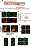
FIGURE 1.
FIGURE 1.
Plasma membrane translocation of LID. A, topology model of NHE1 and amino acid sequence of LID. Positions of two mutant constructs (LI1 and LI2) are represented. Leu-562 and Ile-563 in LI1 and Leu-573 and Ile-574 in LI2 were replaced with two alanine residues. B, PMA (1 μm) facilitates translocation of GFP-labeled LID (GFP-WT-LID) to the plasma membrane, in a similar time course as mCherry-labeled PKCδ-C1a domain. Note that fluorescence signals detected in the nucleus decreased and accumulated in the plasma membrane. C, time courses for the accumulation of two fluorescent probes in the plasma membrane within the same cells. Means ± S.E. (n = 7). D, colocalization efficiency was analyzed for a cell in B before and 10 min after PMA addition. High Pierson's coefficient (0.918) was observed with PMA. E, PMA does not promote the plasma membrane translocation of the 27 N-terminal residues of LID. OAG (100 μm) (F) and phenylephrine (Phe) (1 μm) (G) facilitate plasma membrane translocation of LID. In G, α1AR was stably expressed. H, PMA does not promote plasma membrane translocation of two LID mutants.
FIGURE 2.
FIGURE 2.
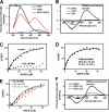
PEs directly interact with LID in vitro. A, emission spectra at 280-nm excitation. FRET signals were observed between a tryptophan residue of peptide GP57 spanning LID (10 μm) and the fluorescent PE analogue SAPD (20 μm). B, difference emission spectra. In peptides GC20 and IL54, FRET signals (increment in fluorescence intensity at 440 nm) were not observed. C, FRET signals were considerably reduced in the presence of high concentrations of PMA (100 μm). D, liposomes increased SAPD affinity (see Table 1). E, PMA (10 μm), OAG (100 μm), and 4α-PMA (100 μm) decrease the apparent affinity of GP57 for SAPD, suggesting that they competitively interact with GP57. F, two mutations, particularly LI2, markedly reduced FRET signals.
FIGURE 3.
FIGURE 3.
Multiple phospholipids can interact with LID. A, CBB-stained patterns of peptide GP57 spanning LID. Liposomes containing various lipids (weight %, adjusted to 100% with PC) were mixed with GP57 and centrifuged. Peptides before centrifugation (total) and an aliquot of each supernatant were subjected to PAGE. It was difficult to obtain the reliable data for interaction of DAG with peptides using this method because DAG inhibited the stable liposome formation. B, relative amount of GP57 bound to liposomes was calculated as described under “Experimental Procedures.” GP57 preferentially binds to acidic phospholipids such as PS, PG, PI, PIP2, and PA. Approximately 50% of peptides also bind to liposomes consisting of only PC under these conditions, suggesting that GP57 is capable of binding various membrane lipids. C, binding of peptides containing replaced residues to PC or PC + PS liposomes. CBB-stained patterns of peptides GP57-LI1 and GP57-LI2 from total input and the supernatant. Summarized data are represented in the lower panel. One amino acid substitution (GP57-LI2) markedly inhibited lipid binding, whereas another substitution (GP57-LI1) had a small inhibitory effect. Means ± S.D. (n = 3–4). *, p < 0.05 versus GP57.
FIGURE 4.
FIGURE 4.
Effect of LID mutation on NHE1 activity and regulation. A, change in pHi was measured using the pHi indicator BCECF-AM. PS120 cells were stably transfected with WT NHE1 (upper), WT + α1AR (lower left), or LI1 + α1AR (lower right). Cells were stimulated with PMA (1 μm), OAG (100 μm), or phenylephrine (Phe) (1 μm). In one experiment, cells were preincubated for 15 min with wortmannin (wort) (10 μm) and then stimulated with phenylephrine in the presence of wortmannin. Means ± S.E. (n > 20 cells). B, change in pHi 15 min after stimulation was measured using the [14C]benzoic acid equilibration method. Means ± S.D. (n = 3). C, SAPD-induced cytoplasmic alkalinization measured in cells expressing WT or mutant NHE1 by using the 14C-benzoic acid equilibration method. This alkalinization was abolished by mutations in LID. Means ± S.D. (n = 3). D, LID mutations induced an acidic shift of pHi dependence (decreased cytosolic H+ affinity).
FIGURE 5.
FIGURE 5.
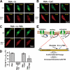
Effect of PKC inhibitors and 4α-PMA on PE-induced NHE1 activation. A, effect of PKC inhibitors on the translocation of myristoylated alanine-rich C kinase substrate (MARCKS) from the plasma membrane to the cytoplasm. Cells transiently expressing the MARCKS-GFP protein were preincubated with PKC inhibitors (1 μm each) for 15 min, and then PMA (1 μm) together with these inhibitors were added at time 0. These PKC inhibitors almost completely abrogated the translocation of MARCKS from the plasma membrane to the cytoplasm, confirming that they inhibited PKC. BIM, bisindolylmaleimide I; CalC, calphostin C; Go, Go-6976; Ro, Ro-32–0432; and St, staurosporin, Scale, 10 μm. B, under conditions similar to those in the experiment of MARCKS translocation, the effect of PKC inhibitors on PMA- or phenylephrine (Phe)-induced NHE1 activation was examined. Except for staurosporin (St), all other PKC inhibitors tested had only a marginal inhibitory effect on NHE1 activation. C, staurosporin, but not calphostin C, decreased cytosolic H+ affinity in WT-NHE1-expressing cells. D, 4α-PMA competitively inhibited PMA-induced cytoplasmic alkalinization. *, p < 0.05; **, p < 0.01.
FIGURE 6.
FIGURE 6.
Differential effects of lipophilic compounds on plasma membrane translocation of LID. A, staurosporin (St) inhibits PMA-induced LID (upper), but not PMA-induced PKCδ-C1a (lower), plasma membrane translocation. B, in contrast, calphostin C (CalC) does not inhibit PMA-induced LID (upper) translocation, but does inhibit PKCδ-C1a (lower) plasma membrane translocation. C, similar to staurosporin, 4α-PMA partly inhibits LID, but not PKCδ-C1a, plasma membrane translocation. D, relative change (%) in the fluorescence intensity of GFP-LID localized in the plasma membrane before and after (10 min) the addition of PMA and/or other reagents. Means ± S.E. (n = 5–7). *, p < 0.05 versus PMA alone. E, schematic models for NHE1 regulation (upper) with LID (lower). In the resting state, physiological NHE1 activity is preserved by interaction of LID with membrane phospholipids. Upon PE or receptor stimulation, PE/DAG binds to LID (probably its C-terminal portion), induces a conformational change to increase its affinity for membrane lipids, and thereby increases H+ affinity, leading to NHE1 activation. Mutations or interaction with some lipophilic compounds such as staurosporin reduce the affinity of LID to lipids, and thus inhibit NHE1 by decreasing H+ affinity.

Similar articles

Cited by

References

    1. Nishizuka Y. (1992) Science 258, 607–614 - PubMed
    1. Griner E. M., Kazanietz M. G. (2007) Nat. Rev. Cancer 7, 281–294 - PubMed
    1. Moolenaar W. H., Tertoolen L. G., de Laat S. W. (1984) Nature 312, 371–374 - PubMed
    1. Grinstein S., Cohen S., Goetz J. D., Rothstein A. (1985) J. Cell Biol. 101, 269–276 - PMC - PubMed
    1. Besterman J. M., Cuatrecasas P. (1984) J. Cell Biol. 99, 340–343 - PMC - PubMed

Publication types

LinkOut - more resources