. 2010 Aug 20;285(34):26199-210.
doi: 10.1074/jbc.M110.103127. Epub 2010 Jun 15.

Erk5 activation elicits a vasoprotective endothelial phenotype via induction of Kruppel-like factor 4 (KLF4)

Affiliations

Erk5 activation elicits a vasoprotective endothelial phenotype via induction of Kruppel-like factor 4 (KLF4)

Nils Ohnesorge et al. J Biol Chem. .

Abstract

The MEK5/Erk5 MAPK cascade has recently been implicated in the regulation of endothelial integrity and represents a candidate pathway mediating the beneficial effects of laminar flow, a major factor preventing vascular dysfunction and disease. Here we expressed a constitutively active mutant of MEK5 (MEK5D) to study the transcriptional and functional responses to Erk5 activation in human primary endothelial cells. We provide evidence that constitutive Erk5 activation elicits an overall protective phenotype characterized by increased apoptosis resistance and a decreased angiogenic, migratory, and inflammatory potential. This is supported by bioinformatic microarray analysis, which uncovered a statistical overrepresentation of corresponding functional clusters as well as a significant induction of anti-thrombotic, hemostatic, and vasodilatory genes. We identify KLF4 as a novel Erk5 target and demonstrate a critical role of this transcription factor downstream of Erk5. We show that KLF4 expression largely reproduces the protective phenotype in endothelial cells, whereas KLF4 siRNA suppresses expression of various Erk5 targets. Additionally, we show that vasoprotective statins potently induce KLF4 and KLF4-dependent gene expression via activation of Erk5. Our data underscore a major protective function of the MEK5/Erk5/KLF4 module in ECs and implicate agonistic Erk5 activation as potential strategy for treatment of vascular diseases.


Figures
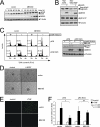
FIGURE 1.
FIGURE 1.
MEK5D expression elicits an anti-apoptotic, anti-angiogenic, and anti-inflammatory phenotype in human EC. A, HUVECs were infected with either an empty retrovirus or a retrovirus encoding MEK5D. At the indicated time points after infection, cells were lysed and analyzed for Erk5 phosphorylation and MEK5D expression by immunoblot using antisera against Erk5 or MEK5, respectively. Tubulin served as the loading control. B, U2OS osteosarcoma cells were co-transfected with pBP-MEK5D and FLAG-wt-Erk5 or MEK5-insensitive FLAG-Erk5-AEF, and phosphorylation and expression of the indicated proteins was detected by immunoblot using antibodies against MEK5, FLAG, phospho-Erk1/2, and tubulin (as loading control). C–F, HUVECs were infected with the indicated retroviruses. Except for D, cells were puromycin-selected and reseeded for the respective assays. C, left panel, shown is flow cytometric quantification of subdiploid DNA content (indicating apoptosis) of PI-stained cells cultured in presence or absence of growth factors for 48 h. Co-expression of Erk5 shRNA (pRS-Erk5) reverses the anti-apoptotic effect of active MEK5D. Right panel, shown is a Western blot, confirming MEK5D protein expression and knockdown efficiency of the Erk5 shRNA construct. An immunoblot for tubulin was included as loading control. D, decreased basal and VEGF-induced angiogenic sprouting of MEK5D-infected HUVEC in a three-dimensional in vitro sprouting assay 24 h after incubation in VEGF-free or VEGF-containing (10 ng/ml) medium. E, a fluorescence microscopy image shows decreased adhesion of fluorochrome-labeled THP-1 monocytes to a monolayer of TNF-stimulated (2 ng/ml, 16 h) ECs upon stable expression of MEK5D. F, quantitative leukocyte adhesion assays show decreased adhesion of fluorescently labeled human THP-1 monocytes to TNF-stimulated ECs expressing MEK5D and reversion by retroviral co-expression of Erk5 shRNA. Adhesion was quantified by measuring fluorescence of total lysates at an appropriate wavelength. Data represent average-fold fluorescence values related to unstimulated pBP/pRS vector co-infected cells (arbitrarily set to 1) and are derived from three independent experiments. ns, not significant.
FIGURE 2.
FIGURE 2.
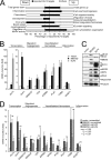
Endothelial targets of active Erk5. A and B, HUVECs were infected with MEK5D or control vector, and RNA was isolated 40 h post-infection. A, qRT-PCRs validating MEK5D-dependent expression of nine selected Erk5 targets from the microarray analysis. Relative expression of the indicated genes was normalized to glyceraldehyde-3-phosphate dehydrogenase expression and subsequently related to empty vector-infected cells (arbitrarily set to 1) to calculate -fold mRNA induction. Data show the means from three independent experiments ± S.D. B, bioinformatic functional annotation cluster analysis of the MEK5D-microarray data using DAVID is shown. The given enrichment score ranks the indicated functional clusters according to statistical overrepresentation of its included GO groups in relation to the totally represented genes on the chip. Only functional annotation clusters containing at least one GO group with statistically significant p value (<0.05) are shown. C–E, HUVEC were infected with empty vector or MEK5D and either lysed 96 h after infection for Western blots for verification of KLF4 protein induction (C) or transfected with the indicated siRNAs before infection and processed for Western blot against the indicated proteins (D) or for qRT-PCR analysis of KLF2 mRNA (E, left) or KLF4 mRNA expression (E, right) 40 h after infection, respectively. ns, not significant.
FIGURE 3.
FIGURE 3.
KLF4 regulates gene expression downstream of Erk5. A, a representation is shown depicting the percentage of reported KLF2 target genes among the MEK5D-regulated functional annotation clusters as identified by comparison of our data set of MEK5D-regulated genes to microarray data of published endothelial KLF2 targets (12). The bars indicate the percentages of reported KLF2 targets (black) or putatively KLF2-independent genes (other, non-filled) calculated for all up- and down-regulated genes (top) or for the individual MEK5D-regulated functional clusters, respectively. The total percentages of reported KLF2 targets for the overall 172 up-regulated/induced genes and 67 repressed transcripts identified by our MEK5D microarray is indicated by dashed lines for comparison. B–D, HUVECs were retrovirally infected as indicated and processed differently for the individual experiments. B, RNA samples were taken 40 h post-infection and subjected to qRT-PCR for quantification of -fold mRNA expression of the indicated genes related to control vector-infected cells. Data display average values from three experiments ±S.D. C, cells were treated overnight with 2 μm monensin to prevent release of extracellular proteins (e.g. ADAMTS1, PAI2, and NOV), lysed 40 h after infection, and analyzed by Western blot using specific antisera against the identified MEK5D target proteins. Tubulin served as loading control. D, HUVECs were transfected with siRNA against KLF2, KLF4, or scrambled siRNA 4 h before infection with empty vector or MEK5D. Expression of the indicated genes was monitored 40 h post-infection by qRT-PCR as described in B. Additionally, in B and D a functional classification of the genes based on published evidence or assignment to specific functional annotation clusters identified by the DAVID algorithm is indicated. ns, not significant.
FIGURE 4.
FIGURE 4.
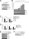
Expression of KLF4 and MEK5D suppresses TNF-induced inflammatory responses by inhibiting NFκB-dependent gene expression at the level of transactivation. HUVECs were retrovirally infected with empty vector, MEK5D, or KLF4, puromycin-selected, and reseeded for the respective experiments. A, qRT-PCR data for VCAM1, demonstrating statistically significant reduction of TNF-induced VCAM1 mRNA upon expression of KLF4 or MEK5D are shown. B, expression of KLF4 and MEK5D does not alter the kinetics of TNF-induced IκBα degradation. Cells were stimulated with TNF for the indicated time points, and total lysates were examined for IκBα protein degradation by Western blot. C, expression of KLF4 or MEK5D inhibits activity of a NFκB-dependent luciferase reporter. The differently infected HUVECs were co-transfected with a NFκB-responsive 6× κB-firefly luciferase and a renilla-luciferase reporter, and luciferase activities were measured 16 h after stimulation with medium or TNF in a luminometer. The data represent -fold ratios of firefly/renilla activities ± S.D. related to unstimulated empty vector-infected cells, which were arbitrarily set to 1. D, enzyme-linked immunosorbent assays (ELISA) show decreased protein levels of the NFκB-dependent proinflammatory cytokines interleukin-8 (IL8) and MCP1 in the culture supernatants of TNF-stimulated HUVEC stably expressing KLF4 or MEK5D, respectively. Data in A, C, and D are derived from 3–5 independent experiments and show average values ±S.D.
FIGURE 5.
FIGURE 5.
KLF4 overexpression mimics the anti-apoptotic and anti-angiogenic effects of active Erk5. HUVECs were retrovirally infected with MEK5D, KLF4, or control vector. Except for C, cells were puromycin-selected 72 h post-infection and reseeded for the individual experiments. A and B, cells were deprived of growth factors and fetal bovine serum for 48 h or incubated in normal growth medium for 48 h. Apoptosis induction was monitored by detection of cleaved caspase-3 (Casp3) using a cleavage-specific antiserum (A) or analyzed by measuring subdiploid DNA content of PI-stained cells by flow cytometry (B). Western blots for MEK5, KLF4, and tubulin in A served as expression or loading controls, respectively. Additionally, an immunoblot for Erk5 is shown to confirm activity of the expressed MEK5D construct. C, shown is decreased basal and VEGF-induced angiogenic sprouting of MEK5D- or KLF4-infected HUVECs as determined in three-dimensional-sprouting assays. Data represent quantifications of the average total sprouting length of each 10 spheroids from three independent experiments ±S.D. The spheroids were seeded into collagen and stimulated with 10 ng/ml VEGF or medium alone for 24 h. D, left, microscopic images show decreased migration capacity of the KLF4- and MEK5D-expressing cells as determined by wounding assay. Images are taken 16 h after infliction of the damage. Right, measurement of the averaged distances of migration ± S.D. obtained for the differently infected cells is shown. Data are averaged from five experiments.
FIGURE 6.
FIGURE 6.
Statins induce KLF4 and KLF4-dependent gene expression via activation of Erk5. HUVECs were treated with each 10 μm concentrations of the indicated statins for 24 h unless indicated otherwise. A, Erk5 phosphorylation and expression of KLF4 and its targets NOS3 and ADAMTS1 were determined by Western blot. Tubulin served as the loading control. B, simvastatin triggers Erk5 phosphorylation and induction of KLF4 mRNA and NOS3 protein in a concentration-dependent manner. Bars show qRT-PCR experiments for KLF4 averaged from three experiments ±S.D. An immunoblot for tubulin is shown to indicate equal loading of the lanes for Erk5 and NOS3, respectively. C, left panel, suppression of simvastatin-induced KLF4 mRNA production upon transfection with siRNA against MEK5 is shown. Data represent the averages of three independent qRT-PCR experiments ±S.D. For control of knockdown levels, the respective qRT-PCRs for MEK5 are shown (right panel). D, qRT-PCR data for KLF4, ADAMTS1, and NOS3 reveal mRNA induction by simvastatin and reversion by expression of shRNA against Erk5. E, depletion of Erk5 prevents simvastatin-induced protein expression of NOS3 and ADAMTS1. F, transfection of siRNA against KLF4 reduces simvastatin-induced NOS3 protein production.

Similar articles

Cited by

References

    1. Traub O., Berk B. C. (1998) Arterioscler. Thromb. Vasc. Biol. 18, 677–685 - PubMed
    1. Hayashi M., Lee J. D. (2004) J. Mol. Med. 82, 800–808 - PubMed
    1. Yan C., Takahashi M., Okuda M., Lee J. D., Berk B. C. (1999) J. Biol. Chem. 274, 143–150 - PubMed
    1. Nishimoto S., Nishida E. (2006) EMBO Rep. 7, 782–786 - PMC - PubMed
    1. Nakamura K., Uhlik M. T., Johnson N. L., Hahn K. M., Johnson G. L. (2006) Mol. Cell. Biol. 26, 2065–2079 - PMC - PubMed

Publication types

MeSH terms