. 2010 Aug 20;285(34):26162-73.
doi: 10.1074/jbc.M109.096453. Epub 2010 Jun 15.

Proteolysis-induced N-terminal ectodomain shedding of the integral membrane glycoprotein CUB domain-containing protein 1 (CDCP1) is accompanied by tyrosine phosphorylation of its C-terminal domain and recruitment of Src and PKCdelta

Affiliations

Proteolysis-induced N-terminal ectodomain shedding of the integral membrane glycoprotein CUB domain-containing protein 1 (CDCP1) is accompanied by tyrosine phosphorylation of its C-terminal domain and recruitment of Src and PKCdelta

Yaowu He et al. J Biol Chem. .

Abstract

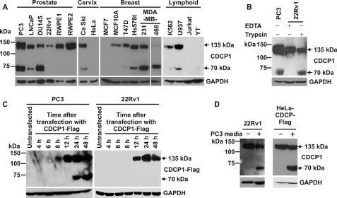
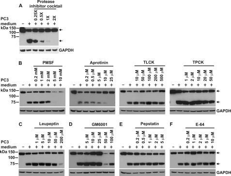
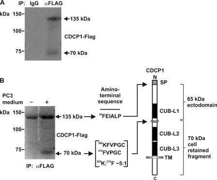
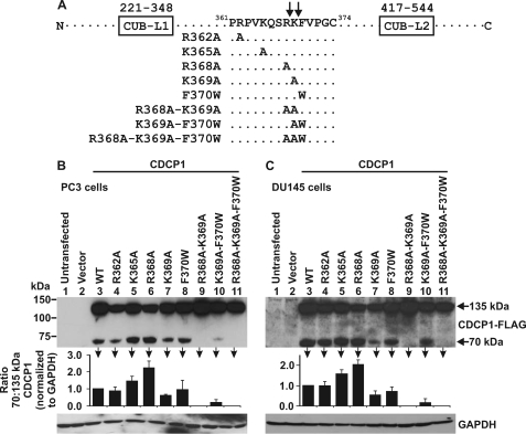
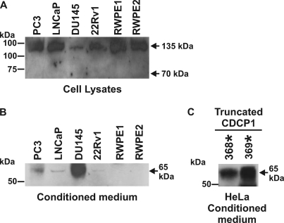
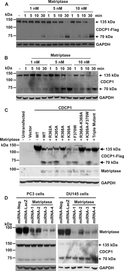
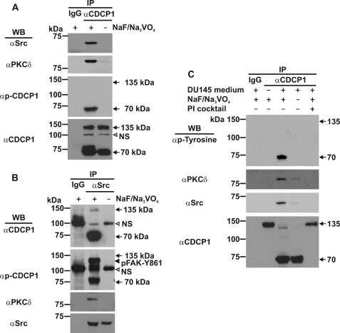
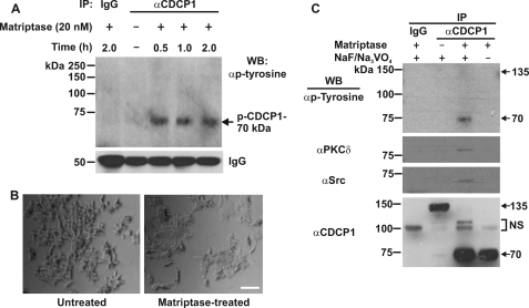
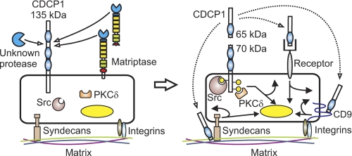
CUB-domain-containing protein 1 (CDCP1) is an integral membrane glycoprotein with potential as a marker and therapeutic target for a number of cancers. Here we examine mechanisms regulating cellular processing of CDCP1. By analyzing cell lines exclusively passaged non-enzymatically and through use of a panel of protease inhibitors, we demonstrate that full-length 135 kDa CDCP1 is post-translationally processed in a range of cell lines by a mechanism involving serine protease activity, generating a C-terminal 70-kDa fragment. Immunopurification and N-terminal sequencing of this cell-retained fragment and detailed mutagenesis, show that proteolytic processing of CDCP1 occurs at two sites, Arg-368 and Lys-369. We show that the serine protease matriptase is an efficient, but not essential, cellular processor of CDCP1 at Arg-368. Importantly, we also demonstrate that proteolysis induces tyrosine phosphorylation of 70-kDa CDCP1 and recruitment of Src and PKCdelta to this fragment. In addition, Western blot and mass spectroscopy analyses show that an N-terminal 65-kDa CDCP1 ectodomain is shed intact from the cell surface. These data provide new insights into mechanisms regulating CDCP1 and suggest that the biological role of this protein and, potentially, its function in cancer, may be mediated by both 70-kDa cell retained and 65-kDa shed fragments, as well as the full-length 135-kDa protein.


Figures

FIGURE 1.
FIGURE 1.
Post-translational processing generates 70 kDa CDCP1 from the full-length 135 kDa protein. A, Western blot analysis using a goat antibody generated against the last 13 C-terminal residues of CDCP1 (Abcam ab1377) was performed on lysates from the indicated cell lines passaged with EDTA. B, trypsin induces the generation of 70 kDa CDCP1. Lysates from prostate cancer PC3 cells passaged with EDTA and 22Rv1 cells treated once with either EDTA or trypsin (5 min) were examined by Western blot analysis using a goat anti-CDCP1 antibody (Abcam ab1377). C, prostate cancer PC3 and 22Rv1 cells were transiently transfected with a CDCP1-Flag expression construct, and lysates were collected at the indicated time points for anti-Flag Western blot analysis. D, Western blot analysis of lysates from 22Rv1 (left panel) and HeLa-CDCP1-Flag (right panel) cells either untreated or cultured for 24 h in PC3 cell-conditioned medium. Lysates from 22Rv1 cells were probed with a goat anti-CDCP1 antibody (Abcam ab1377) and lysates from HeLa-CDCP1-Flag cells were probed with a rabbit anti-Flag antibody. Anti-GAPDH Western blot analysis was performed to examine protein loading.
FIGURE 2.
FIGURE 2.
Serine protease activity is required for conversion of 135 kDa CDCP1 to 70 kDa. Anti-Flag Western blot analysis of lysates from HeLa-CDCP1-Flag cells untreated or cultured for 24 h in PC3 cell-conditioned medium that was either untreated or had been supplemented with (A) Complete EDTA-free protease inhibitor mixture or (B) PMSF, aprotinin, TLCK, or TPCK, (C) leupeptinin, (D) GM6001, (E) pepstatin, or (F) E-64 at the indicated concentrations. Arrows indicate 70 and 135 kDa CDCP1. Anti-GAPDH Western blot analysis was performed to examine protein loading.
FIGURE 3.
FIGURE 3.
Identification of the sites at which CDCP1 is proteolytically processed. A, anti-Flag Western blot analysis of proteins obtained from mouse IgG and anti-Flag antibody immunoprecipitations from lysates of HeLa-CDCP1-Flag cells treated with PC3 cell-conditioned medium. B, Coomassie-stained PVDF membrane of anti-Flag immunoprecipitates obtained from HeLa-CDCP1-Flag cells either untreated or treated with PC3 cell-conditioned medium. To the right is shown the N-terminal sequence obtained by Edman degradation sequencing of 70 and 135 kDa CDCP1 and a diagram showing the location of the identified N termini within the CDCP1 structure. The data indicated that 135 kDa CDCP1 is processed after Arg-368 and Lys-369 in the ratio 5:1 in a region located between CUB-like domains 1 and 2 (CUB-L1 and CUB-L2). SP, signal peptide; TM, transmembrane domain.
FIGURE 4.
FIGURE 4.
Examination of the cellular proteolytic processing sites of CDCP1. A, schematic showing the location of single, double, and triple amino acid mutations introduced into the CDCP1-Flag sequence between CUB-like domain 1 and 2 (CUB-L1 and CUB-L2), R362A, K365A, R368A, K369A, F370W, R368A-K369A, K369A-F370W, and R368A-K369A-F370W. B, PC3 and C, DU145 cell lysates from cells either untransfected or transiently transfected with either vector or wild type or mutant CDCP1-Flag expression constructs analyzed by anti-Flag Western blot analysis. Anti-GAPDH Western blot analysis was performed to examine protein loading. Graphs were generated from densitometric analysis of at least three separate experiments. For 70 and 135 kDa CDCP1 and GAPDH, signal intensities from cells transfected with mutant constructs were normalized against the signal from cells transfected with wild-type CDCP1. These normalized values were used to calculate the ratio of 70 to 135 kDa CDCP1.
FIGURE 5.
FIGURE 5.
The CDCP1 ectodomain is shed intact from the cell surface. Western blot analyses using a goat antibody generated against the extracellular domain of CDCP1 (R&D Systems AF2666). A, analysis of lysates from prostate cell lines passaged with EDTA demonstrating that this antibody recognizes 135 kDa but not 70 kDa CDCP1 indicating that the cognate antigen is located in the shed portion of CDCP1. B, analysis of conditioned media from these cell lines. Cells were cultured in serum-free medium for 3 days. The medium was then centrifuged briefly to remove intact cells, and proteins were recovered by acetone precipitation. C, analysis of serum-free conditioned media from HeLa cells transiently transfected with expression constructs encoding CDCP1 truncated after either Arg-368 (designated R368*) or Lys-369 (K370*).
FIGURE 6.
FIGURE 6.
The serine protease matriptase is an efficient but not essential processor of CDCP1. A, anti-Flag Western blot analysis of lysates from HeLa-CDCP1-Flag cells either untreated (−) or incubated with 1, 5, or 10 nm matriptase for 1, 5, 10 or 30 min. B, Western blot analysis of lysates from 22Rv1 cells either untreated (−) or incubated with 1, 5, or 10 nm matriptase for 1, 5, 10, or 30 min using a goat anti-CDCP1 antibody (Abcam ab1377). Anti-GAPDH Western blot analysis was performed to examine protein loading. C, HeLa cells were either untransfected, transfected with vector alone, or co-transfected with matriptase and either wild type (WT) CDCP1-Flag expression construct or constructs encoding the indicated single and double residue mutant CDCP1-Flag constructs or triple mutant CDCP1-Flag-R368A-K369A-F370W. After 24 h, lysates were collected and analyzed by anti-Flag, anti-matriptase, and anti-GAPDH Western blot analysis. D, polyclonal populations of PC3 and DU145 cells were stably transfected with one of four CDCP1 knockdown miRNA constructs. After 4 weeks of selection in blasticidin, cell lysates were analyzed by Western blot analysis using rabbit anti-matriptase, goat anti-CDCP1 (Abcam ab1377), and anti-GAPDH antibodies.
FIGURE 7.
FIGURE 7.
Examination of tyrosine phosphorylation of CDCP1 and binding of Src and PKCδ induced by DU145 media. A, anti-Src, -PKCδ, -p-CDCP1-Y734 (performed with an antibody that detects both p-CDCP1-Y734 and p-FAK-Y861), and -CDCP1 (CST 4115) Western blot analysis of anti-CDCP1 immunoprecipitates (CST 4115) obtained from DU145 cells. B, anti-CDCP1 (CST 4115), -p-CDCP1-Y734 (performed with an antibody that detects both p-CDCP1-Y734 and p-FAK-Y861), -PKCδ, and -Src Western blot analysis of anti-Src immunoprecipitates obtained from DU145 cells. C, anti-phosphotyrosine, anti-PKCδ, anti-Src, and anti-CDCP1 (CST 4115) Western blot analysis of anti-CDCP1 immunoprecipitates obtained from 22Rv1 cells either untreated (−) or treated (+) with 3 day serum-free conditioned medium from DU145 cells for 36 h. The media was either untreated (−) or treated (+) with protease inhibitor (PI) mixture before incubation with 22Rv1 cells. Lysates from all experiments were collected in the presence (+) or absence (−) of sodium vanadate and sodium fluoride. All experiments included control immunoprecipitations performed with species matched IgG. NS, nonspecific.
FIGURE 8.
FIGURE 8.
Matriptase proteolysis mediates tyrosine phosphorylation and binding of Src and PKCδ to 70 kDa CDCP1. A, anti-phosphotyrosine Western blot analysis of proteins immunoprecipitated from 22Rv1 cell lysates using a rabbit anti-CDCP1 antibody (CST 4115). Lysates were collected from 22Rv1 cells either untreated (−) or treated (+) with 20 nm matriptase for the indicated times. The blot was reprobed with an anti-rabbit secondary antibody to detect rabbit IgG to assess consistency in the amount of antibody used for immunoprecipitations. B, photographic images of 22Rv1 cells either untreated (left panel) or treated for 2 h with 20 nm matriptase (right panel). Bar, 50 μm. C, anti-phosphotyrosine, -PKCδ, -Src, and -CDCP1 (CST 4115) Western blot analysis of anti-CDCP1 (CST 4115) immunoprecipitates obtained from 22Rv1 cells either untreated (−) or treated (+) with matriptase (20 nm; 0.5 h). Lysates were collected in the presence (+) or absence (−) of sodium vanadate and sodium fluoride. All experiments included control immunoprecipitations performed with species matched IgG. NS, nonspecific.
FIGURE 9.
FIGURE 9.
Protease-mediated processing of CDCP1. Serine proteolysis generates a 65 kDa shed ectodomain and a tyrosine-phosphorylated cell retained 70 kDa CDCP1 fragment. Proteolysis occurs at Arg-368 and Lys-369 between CUB-like domain 1 and 2 of CDCP1 (the 3 CDCP1 CUB-like domains are shown as blue ovals). Proteolysis results in tyrosine phosphorylation of 70 kDa CDCP1 (yellow-filled circles) and recruitment of Src and PKCδ in a phosphorylation-dependent manner. We propose that the CDCP1 ectodomain may function as a ligand or competitive inhibitor for a cell surface receptor (autocrine receptor binding is shown, but paracrine and endocrine signaling may also be relevant) or as a matrix-interacting protein potentially modulating cell:matrix interactions occurring via known CDCP1 interacting proteins such as syndecan 1 and 4 and the tetraspannin CD9 (dotted arrows). Potential signaling downstream of the 70 kDa CDCP1 cell-retained fragment and ectodomain resulting in direct cellular responses and changes in gene expression are represented by curved and straight arrows, respectively. Currently the endogenous serine protease or proteases mediating cleavage of CDCP1 at Arg-368 and Lys-369 are not known. However, we have shown that the cell surface and shed serine protease matriptase efficiently cleaves CDCP1 exclusively at Arg-368.

Similar articles

Cited by

References

    1. Scherl-Mostageer M., Sommergruber W., Abseher R., Hauptmann R., Ambros P., Schweifer N. (2001) Oncogene 20, 4402–4408 - PubMed
    1. Hooper J. D., Zijlstra A., Aimes R. T., Liang H., Claassen G. F., Tarin D., Testa J. E., Quigley J. P. (2003) Oncogene 22, 1783–1794 - PubMed
    1. Brown T. A., Yang T. M., Zaitsevskaia T., Xia Y., Dunn C. A., Sigle R. O., Knudsen B., Carter W. G. (2004) J. Biol. Chem. 279, 14772–14783 - PubMed
    1. Bhatt A. S., Erdjument-Bromage H., Tempst P., Craik C. S., Moasser M. M. (2005) Oncogene 24, 5333–5343 - PMC - PubMed
    1. Bühring H. J., Kuçi S., Conze T., Rathke G., Bartolović K., Grünebach F., Scherl-Mostageer M., Brümmendorf T. H., Schweifer N., Lammers R. (2004) Stem. Cells 22, 334–343 - PubMed

Publication types