. 2010 Aug 20;285(34):26269-78.
doi: 10.1074/jbc.M109.095752. Epub 2010 Jun 15.

Calcineurin-mediated dephosphorylation of synaptotagmin VI is necessary for acrosomal exocytosis

Affiliations

Calcineurin-mediated dephosphorylation of synaptotagmin VI is necessary for acrosomal exocytosis

Jimena Castillo Bennett et al. J Biol Chem. .

Abstract

Regulated secretion is a fundamental process underlying the function of many cell types. In particular, acrosomal exocytosis in mammalian sperm is essential for egg fertilization. In general, exocytosis is initiated by a cytosolic calcium increase. In this report we show that calcium affects several factors during human sperm acrosomal exocytosis. By using an antibody that specifically recognizes synaptotagmin VI phosphorylated at the polybasic region of the C2B domain, we showed that a calcium-dependent dephosphorylation of this protein occurred at early stages of the acrosomal exocytosis in streptolysin O-permeabilized sperm. We identified the phosphatase as calcineurin and showed that the activity of this enzyme is absolutely required during the early steps of the secretory process. When added to sperm, an inhibitor-insensitive, catalytically active domain of calcineurin was able to rescue the effect of the specific calcineurin inhibitor cyclosporin A. This same domain dephosphorylated recombinant synaptotagmin VI C2B domain, validating this protein as a new substrate for calcineurin. When sperm were treated with catalytically active calcineurin before stimulation, exocytosis was inhibited, an effect that was rescued by the phosphomimetic synaptotagmin VI C2B-T418E,T419E mutant domain. These observations indicate that synaptotagmin must be dephosphorylated at a specific window of time and suggest that phosphorylated synaptotagmin has an active role at early stages of the acrosomal exocytosis.


Figures
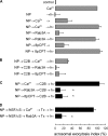
FIGURE 1.
FIGURE 1.
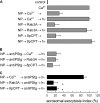
Role of calcium during early stages of acrosomal exocytosis. Permeabilized spermatozoa were loaded with 10 μm NP-EGTA (NP) for 15 min at 37 °C to chelate intra-acrosomal Ca2+. Acrosomal exocytosis (AE) was then initiated by adding 10 μm free Ca2+, 300 nm Rab3A, or 50 μm 8pCPT. After a further 10 min of incubation at 37 °C to allow exocytosis to proceed up to the intra-acrosomal Ca2+-sensitive step, sperm were treated for 10 min at 37 °C with 500 nm synaptotagmin VI C2B domain (C2B). All of these procedures were carried out in the dark. UV flash photolysis of the chelator was induced at the end of the incubation period (hν), and the samples were incubated for 5 min to promote exocytosis. Relevant experimental conditions are shown as black bars in C. Control experimental conditions shown in A (gray bars) include background AE in the absence of any stimulation (control); AE stimulated by 10 μm free Ca2+; no effect of the combination of NP and light; inhibitory effect of NP-EGTA in the dark; and the recovery upon illumination when AE was initiated with Ca2+, Rab3A, or 8pCPT. Control conditions in B show the inhibitory effect of the C2B domain when included from the beginning of the experiment. In D, after loading the acrosome with NP-EGTA, the sperm were stimulated in the presence of 310 nm NSF plus 500 nm αSNAP (NSFS) with Ca2+ or Rab3A, and then 100 nm tetanus toxin (Tx) was added to cleave SNARE (specifically VAMP) molecules not assembled in SNARE complexes. UV flash photolysis of the chelator was induced at the end of the incubation period (hν), and the samples were incubated for 5 min to promote exocytosis. The sperm were fixed, and AE was measured. The percentage of reacted sperm was normalized as described under “Experimental Procedures.” The data represent the means ± S.E. of at least three independent experiments. The asterisks indicate significant differences from similar conditions stimulated with Ca2+ (p < 0.01, one-way ANOVA and Dunnett test).
FIGURE 2.
FIGURE 2.
Calcium affects synaptotagmin phosphorylation during early stages of acrosomal exocytosis. Permeabilized spermatozoa were loaded with 10 μm NP-EGTA (NP) for 15 min at 37 °C to chelate intra-acrosomal Ca2+. AE was then initiated by adding 10 μm free Ca2+, 300 nm of Rab3A, or 50 μm 8pCPT. After a further 10 min of incubation at 37 °C to allow exocytosis to proceed up to the intra-acrosomal Ca2+-sensitive step, the sperm were treated for 10 min at 37 °C with 70 nm anti-phosphosynaptotagmin antibody (antiPStg). All of these procedures were carried out in the dark. UV flash photolysis of the chelator was induced at the end of the incubation period (hν), and the samples were incubated for 5 min to promote exocytosis. The relevant experimental conditions are shown as black bars in C. Control experimental conditions shown in A (gray bars) include background AE in the absence of any stimulation (control); AE stimulated by 10 μm free Ca2+; inhibitory effect of NP-EGTA in the dark; and the recovery upon illumination when AE was initiated with Ca2+, Rab3A, or 8pCPT. Control conditions in B show the inhibitory effect of the antiPStg when included from the beginning of the experiment. The sperm were fixed, and AE was measured. The percentage of reacted sperm was normalized as described under “Experimental Procedures.” The data represent the means ± S.E. of at least three independent experiments. The asterisks indicate significant differences from similar conditions stimulated with Ca2+ (p < 0.01, one-way ANOVA and Dunnett test).
FIGURE 3.
FIGURE 3.
Calcineurin participates in acrosomal exocytosis. A, a post nuclear extract from rat brain (1 μg of proteins, brain) or human sperm (106 cells, sperm) were resolved in 12.5% gels, transferred to nitrocellulose membranes, and probed with an anti-calcineurin antibody. The molecular mass standard is indicated on the left. B, permeabilized spermatozoa were treated at 37 °C for 10 min with 2 μm CsA, 1 μm FK 506, 67 nm anti-calcineurin antibody (antiCaN), or 100 μm VIVIT for 10 min at 37 °C. AE was then initiated by adding 10 μm free Ca2+, and the incubation continued for an additional 15 min (black bars). Controls include (gray bars) background AE in the absence of any stimulation (control) and AE stimulated by 10 μm free Ca2+. The sperm were fixed, and AE was measured. The percentage of reacted sperm was normalized as described under “Experimental Procedures.” The data represent the means ± S.E. of at least three independent experiments. The asterisks indicate significant differences from 100 (one-way ANOVA and 95% confidence interval for each condition).
FIGURE 4.
FIGURE 4.
Calcium activates a calcineurin-dependent process during the early stages of exocytosis. Permeabilized spermatozoa were loaded with 10 μm NP-EGTA (NP) for 15 min at 37 °C to chelate intra-acrosomal Ca2+. AE was then initiated by adding 10 μm free Ca2+ or 300 nm of Rab3A. After a further 10 min of incubation at 37 °C to allow exocytosis to proceed up to the intra-acrosomal Ca2+-sensitive step, the sperm were treated for 10 min at 37 °C with 2 μm CsA. All of these procedures were carried out in the dark. UV flash photolysis of the chelator was induced at the end of the incubation period (hν), and the samples were incubated for 5 min to promote exocytosis. Relevant experimental conditions are shown as black bars in C. Control experimental conditions shown in A (gray bars) include background AE in the absence of any stimulation (control); AE stimulated by 10 μm free Ca2+; inhibitory effect of NP-EGTA in the dark; and the recovery upon illumination when AE was initiated with Ca2+ or Rab3A. Control conditions in B show the inhibitory effect of the CsA when added before stimulation. The sperm were fixed, and AE was measured. The percentage of reacted sperm was normalized as described under “Experimental Procedures.” The data represent the means ± S.E. of at least three independent experiments. The asterisk indicates significant differences from a similar condition stimulated with Ca2+ (p < 0.01 one-way ANOVA and Dunnett test).
FIGURE 5.
FIGURE 5.
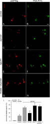
Synaptotagmin dephosphorylation depends on calcium and calcineurin. Nonpermeabilized sperm were incubated for 15 min at 37 °C with 100 μm 2-APB and, when indicated, 1 μm FK 506. The cells were then further incubated for 30 min at 37 °C with no additions (control, A and B) or stimulated with 10 μm A23187 (C, D, G, and H) or 50 μm 8pCPT (E and F). The cells were then fixed and double-stained with the antiPStg antibody followed by an anti-rabbit Cy3 (A, C, E, and G) and FITC-coupled P. sativum agglutinin to differentiate between reacted and intact sperm (B, D, F, and H). The asterisk in F shows a reacted sperm with equatorial lectin staining and very faint antiPStg labeling. Bar, 5 μm. In I, at least 200 cells treated as described for A–H were classified as having or lacking distinct acrosomal phosphosynaptotagmin staining. The percentage of immunolabeled sperm in five independent experiments was recorded. As controls, the percentages of labeled cells when the antiPStg antibody was excluded or when it was preblocked with excess phosphorylated peptide are shown (n = 2, supplemental Fig. S3). The data represent the means ± S.E. or the means ± range. The asterisks indicate significant differences from control sperm incubated with unblocked antiPStg antibody (p < 0.01, one-way ANOVA and Dunnett test).
FIGURE 6.
FIGURE 6.
Synaptotagmin is a substrate for calcineurin. A–C, GST-C2B domains immobilized in glutathione-Sepharose beads were incubated for 40 min at 37 °C with PKCβII under activating conditions in the presence of [γ-32P]ATP. The beads were washed and then incubated for 1 h at 30 °C in the presence or absence of the constitutively active catalytic domain of calcineurin (CA-CaN) in a buffer containing 1 mm MnCl2 (MnCl2 +) or 1 mm EGTA (MnCl2 −). In some experiments, increasing concentrations of an antibody that recognizes the phosphorylated polybasic region of the C2B synaptotagmin domain were added (antiPStg, 0–2.5 μm). The samples were then resolved by SDS-PAGE. Total proteins (top panels) are shown by Coomassie Blue stain. Phosphorylated proteins were detected by autoradiography (bottom panels). D, autoradiographies from three experiments as shown in B were quantified and normalized for the protein load. The data represent the means ± S.E. The asterisk indicates a significant difference from incubation without CA-CaN (one-way ANOVA and 99% confidence interval). E, autoradiographies from two experiments as shown in C were quantified. The data represent the means ± range.
FIGURE 7.
FIGURE 7.
Phosphorylated synaptotagmin has an active role at early stages of the secretory process. A, permeabilized spermatozoa were treated with 2 μm CsA and then stimulated with 10 μm free Ca2+ for 15 min at 37 °C to allow exocytosis to proceed to the calcineurin-dependent step, and finally the inhibition of CsA was relieved by adding 50 nm CA-CaN (black bar). Control experimental conditions (gray bars) include background AE in the absence of any stimulation (control), AE stimulated by 10 μm free Ca2+, and the inhibitory effect of CsA. Two conditions, initially thought as controls, gave unexpected results. When sperm were treated with CA-CaN before activating with calcium (irrespective of the presence of CsA), exocytosis was inhibited (white bars). B, permeabilized spermatozoa were treated with 50 nm CA-CaN for 15 min at 37 °C, then 500 nm C2BTE were added, and finally AE was stimulated with 10 μm free Ca2+ for 15 min at 37 °C (black bar). Control experimental conditions (gray bars) include background AE in the absence of any stimulation (control), AE stimulated by 10 μm free Ca2+, the inhibitory effect of CA-CaN when added before stimulation, and the lack of effect of C2BTE in calcium-triggered exocytosis. C, permeabilized spermatozoa were treated with 500 nm wild type C2B domain in the presence of increasing concentrations of phosphorylated C2B domain (C2BP, left panel) or the phosphomimetic C2B domain (C2BTE, right panel) and stimulated with 10 μm free Ca2+ for 15 min at 37 °C. AE in the presence of 500 nm C2BP or C2BTE is shown as gray bars. The sperm were fixed, and AE was measured. The percentage of reacted sperm was normalized as described under “Experimental Procedures.” The data represent the means ± S.E. of at least three independent experiments. The asterisks indicate significant differences from 100 (one-way ANOVA and 95% confidence interval for each condition).
FIGURE 8.
FIGURE 8.
Working model for the Ca2+/calcineurin-dependent dephosphorylation of synaptotagmin during acrosomal exocytosis. In resting sperm, SNAREs are assembled in inactive cis-complexes, and synaptotagmin is phosphorylated (a). Upon activation, Ca2+ coming from the extracellular medium triggers SNARE complex disassembly and calcineurin-dependent synaptotagmin dephosphorylation. Acrosomal swelling and deformation of the granule membrane favor the tethering with the plasma membrane. SNAREs can then reassemble in trans in combination with complexin; dephosphorylated synaptotagmin is incorporated into the prefusion complex probably interacting with phospholipids in the opposite membrane through the polybasic region (b). We speculate that the calcium concentration at this stage is not sufficient to insert the aspartic-rich Ca2+-binding loops of synaptotagmin into the membrane and to relieve the complexin clamp. These events must wait for the local influx of calcium coming from the acrosome to promote full SNARE complex assembly and membrane fusion (c). When sperm are activated at low calcium concentrations (e.g. with the cAMP analog 8pCPT), the system progresses to the stage where loose trans-complexes are assembled, but synaptotagmin remains phosphorylated (d). The influx of calcium from the acrosome is sufficient to dephosphorylated synaptotagmin at this stage, and the membrane fusion process is completed (c). Unexpectedly, when constitutively active calcineurin is added to resting sperm at low calcium concentrations, synaptotagmin is dephosphorylated, and exocytosis is inhibited (e). The protein may be engaged in unproductive cis-interactions that prevent exocytosis.

Similar articles

Cited by

References

    1. Barclay J. W., Morgan A., Burgoyne R. D. (2005) Cell Calcium 38, 343–353 - PubMed
    1. Burgoyne R. D., Morgan A. (2003) Physiol. Rev. 83, 581–632 - PubMed
    1. Südhof T. C., Rothman J. E. (2009) Science. 323, 474–477 - PMC - PubMed
    1. Mayorga L. S., Tomes C. N., Belmonte S. A. (2007) IUBMB Life 59, 286–292 - PubMed
    1. Yanagimachi R. (1994) in The Physiology of Reproduction (Knobil E., Neill J. D. eds) pp. 189–317, Raven Press, New York

Publication types

LinkOut - more resources