. 2010 Jun;22(6):1733-48.
doi: 10.1105/tpc.109.073072. Epub 2010 Jun 15.

MOTHER OF FT AND TFL1 regulates seed germination through a negative feedback loop modulating ABA signaling in Arabidopsis

Affiliations

MOTHER OF FT AND TFL1 regulates seed germination through a negative feedback loop modulating ABA signaling in Arabidopsis

Wanyan Xi et al. Plant Cell. 2010 Jun.

Abstract

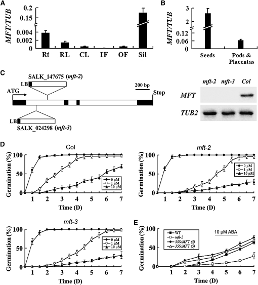
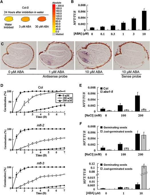
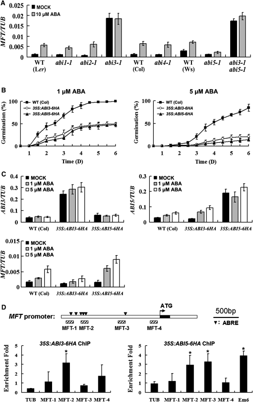
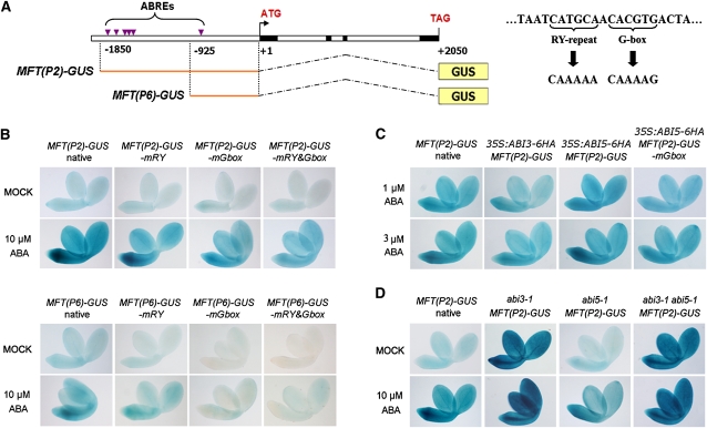
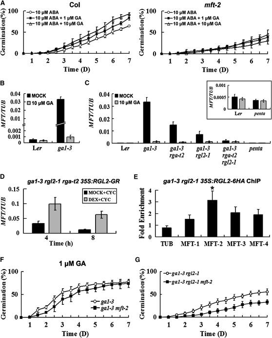
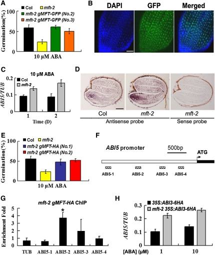
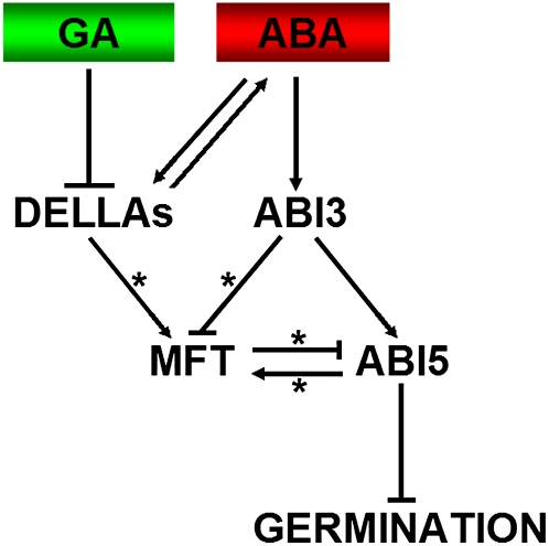
Abscisic acid (ABA) and gibberellin (GA) are two antagonistic phytohormones that regulate seed germination in response to biotic and abiotic environmental stresses. We demonstrate here that MOTHER OF FT AND TFL1 (MFT), which encodes a phosphatidylethanolamine-binding protein, regulates seed germination via the ABA and GA signaling pathways in Arabidopsis thaliana. MFT is specifically induced in the radical-hypocotyl transition zone of the embryo in response to ABA, and mft loss-of-function mutants show hypersensitivity to ABA in seed germination. In germinating seeds, MFT expression is directly regulated by ABA-INSENSITIVE3 (ABI3) and ABI5, two key transcription factors in ABA signaling pathway. MFT is also upregulated by DELLA proteins in the GA signaling pathway. MFT in turn provides negative feedback regulation of ABA signaling by directly repressing ABI5. We conclude that during seed germination, MFT promotes embryo growth by constituting a negative feedback loop in the ABA signaling pathway.


Figures

Figure 1.
Figure 1.
Phenotypic Characterization of mft Mutants in Arabidopsis. (A) Quantitative real-time PCR analysis of MFT expression in various tissues. Results were normalized against the expression of TUB2. Rt, roots; RL, rosette leaves; CL, cauline leaves; IF, inflorescences without open flowers; OF, open flowers; Sil, siliques. Error bars denote sd. (B) MFT expression determined by quantitative real-time PCR in developing siliques dissected into seeds and pods plus placentas. Error bars denote sd. (C) Schematic diagram indicating the T-DNA insertions in two mft loss-of-function mutants, mft-2 (SALK_147675) and mft-3 (SALK_024298). Black and white boxes indicate exons and introns of MFT, respectively. RT-PCR analysis using a pair of primers flanking the T-DNA insertion sites did not detect MFT expression in mft-2 and mft-3, indicating that both of them are null alleles. (D) Germination phenotype of the wild type, mft-2, and mft-3 treated with different concentrations of ABA (0, 1, and 10 μM). Error bars denote sd. (E) Germination phenotype of two representative 35S:MFT lines (3 and 5) in response to 10 μM ABA. Error bars denote sd.
Figure 2.
Figure 2.
MFT Expression Is Upregulated in Response to ABA. (A) Public microarray data showing upregulation of MFT by ABA in germinating seeds (http://www.bar.utoronto.ca/efp/cgi-bin/efpWeb.cgi). The colors from yellow to red indicate the increased absolute signal values of MFT expression retrieved from microarray data. (B) MFT expression determined by quantitative real-time PCR in germinating seeds treated with different concentrations of ABA. All seeds were collected 16 h after stratification. Error bars denote sd. (C) In situ localization of MFT in germinating seeds treated with different concentrations of ABA. Seeds without ABA treatment were collected 12 h after stratification, while other seeds treated with 1 and 10 μM ABA were collected 24 h after stratification. These seeds at the same developmental stage were hybridized with the antisense or sense MFT probe as indicated. Bar = 100 μm. (D) Germination phenotype of the wild type, mft-2, and mft-3 treated with different concentrations of NaCl. Error bars denote sd. (E) MFT expression determined by quantitative real-time PCR in wild-type and aba1-5 germinating seeds treated with different concentrations of NaCl. All seeds were collected 16 h after stratification. Error bars denote sd. (F) MFT expression determined by quantitative real-time PCR in germinating and just-germinated seeds treated with NaCl and ABA. All germinating seeds were collected 16 h after stratification. Seeds with visible protrusion of the radicle tip through all the covering layers were collected as just-germinated seeds. Error bars denote sd.
Figure 3.
Figure 3.
Regulation of MFT by ABA Is Mediated by ABI3 and ABI5. (A) MFT expression determined by quantitative real-time PCR in wild-type (WT) and various abi mutants mock treated or treated with 10 μM ABA. Because abi3-1 seeds germinated around 14 h after stratification, we collected all germinating seeds 12 h after stratification for comparing MFT expression. Error bars denote sd. (B) Germination phenotype of the wild type, 35S:ABI3-6HA, and 35S:ABI5-6HA treated with 1 or 5 μM ABA. Error bars denote sd. (C) Expression of ABI3, ABI5, and MFT determined by quantitative real-time PCR in germinating seeds of the wild type, 35S:ABI3-6HA, and 35S:ABI5-6HA mock treated or treated with 1 or 5 μM ABA. All germinating seeds were collected 16 h after stratification. Error bars denote sd. (D) ChIP enrichment test showing the binding of ABI3-6HA and ABI5-6HA to the MFT promoter. The upstream region and the first intron of MFT are represented by white boxes, while the first exon is represented by a black box. The arrowheads in the top panel indicate the sites containing putative ABREs on the MFT promoter. Hatched boxes represent the DNA fragments amplified in ChIP assays. ChIP assay results of 35S:ABI3-6HA and 35S:ABI5-6HA are shown in the bottom panels. Seeds were sown on Murashige and Skoog (MS) medium supplemented with 10 μM ABA and harvested 16 h after stratification for ChIP assays. Em6, which has been identified as a direct target of ABI5 (Lopez-Molina et al., 2002), is used as a positive control for ABI5-6HA ChIP assay. Significant differences in comparison with the enrichment of a TUB2 fragment are indicated with asterisks (P < 0.05, Student's t test). Error bars denote sd.
Figure 4.
Figure 4.
A G-Box Motif in MFT Promoter Mediates Spatial Regulation of MFT in Response to ABA. (A) Schematic diagram of MFT(P2)-GUS and MFT(P6)-GUS constructs where MFT 5′ upstream sequences containing ABREs were transcriptionally fused with the GUS gene (left panel). The right panel shows the mutagenesis of the RY repeat and the G-box in the ABRE that is located 700 bp upstream of the ATG start codon. (B) GUS staining in germinating seeds of the transformants containing MFT(P2)-GUS, MFT(P6)-GUS, and their derived constructs with the mutated RY repeat (mRY) and G-box motif (mGbox). Seeds from T3 homozygous plants with a single insertion of the transgene for each construct were analyzed, and representative images are shown. Germinating seeds mock treated or treated with 10 μM ABA, which were at the same developmental stage, were stained 12 or 24 h after stratification, respectively. (C) GUS staining in germinating seeds of MFT(P2)-GUS, 35S:ABI3-6HA MFT(P2)-GUS, 35S:ABI5-6HA MFT(P2)-GUS, and 35S:ABI5-6HA MFT(P2)-GUS-mGbox. Germinating seeds treated with 1 and 3 μM ABA, which were at the same developmental stage, were stained 24 h after stratification. (D) GUS staining in germinating seeds of MFT(P2)-GUS, abi3-1 MFT(P2)-GUS, abi5-1 MFT(P2)-GUS, and abi3-1 abi5-1 MFT(P2)-GUS. To examine GUS expression in seeds at the same developmental stage, MFT(P2)-GUS and abi5-1 MFT(P2)-GUS treated with 10 μM ABA were stained 24 h after stratification, while other germinating seeds mock treated or treated with 10 μM ABA were stained 12 h after stratification.
Figure 5.
Figure 5.
Regulation of MFT by GA Is Mediated by DELLA Proteins. (A) Germination phenotype of the wild type and mft-2 treated with 10 μM ABA plus different concentrations of GA. Error bars denote sd. (B) MFT expression determined by quantitative real-time PCR in wild-type and ga1-3 seeds mock treated or treated with 10 μM GA. All germinating seeds were collected 16 h after stratification. Error bars denote sd. (C) MFT expression determined by quantitative real-time PCR in wild-type and various DELLA mutant seeds in ga1-3 background mock treated or treated with 10 μM GA. All germinating seeds were collected 16 h after stratification. penta indicates the ga1-3 gai-t6 rga-t2 rgl1-1 rgl2-1 mutant. Inset shows the comparison of MFT expression between the wild type and penta mutants. Error bars denote sd. (D) MFT expression determined by quantitative real-time PCR in ga1-3 rgl2-1 rga-t2 35S:RGL2-GR seeds. Seeds were treated with 30 μM DEX plus 30 μM cycloheximide (CYC) or MOCK (0.09% ethanol) plus cycloheximide under vacuum for 1 h. They were subsequently washed three times and collected 4 and 8 h after sowing on MS medium. Error bars denote sd. (E) ChIP results showing the binding of RGL2-6HA to the MFT promoter. Primers used for the enrichment test are described in Figure 3D. Seeds were sown on MS medium and harvested 16 h after stratification for ChIP assays. A significant difference in comparison with the enrichment of a TUB2 fragment is indicated with an asterisk (P < 0.05, Student's t test). Error bars denote sd. (F) Germination phenotype of ga1-3 and ga1-3 mft-2 treated with 1 μM GA. Error bars denote sd. (G) Germination phenotype of ga1-3 rgl2-1 and ga1-3 rgl2-1 mft-2 in the absence of exogenous GA. Error bars denote sd.
Figure 6.
Figure 6.
MFT Antagonizes ABA Signaling by Directly Repressing ABI5 during Seed Germination. (A) Two representative mft-2 gMFT-GFP transgenic lines showing rescued germination phenotype in response to 10 μM ABA. The percentage of germination was scored 7 d after stratification. Error bars denote sd. (B) MFT-GFP localization in the cells of the radical-hypocotyl transition zone. DAPI, fluorescence of 4',6-diamidino-2-phenylindole; Merged, merge of DAPI and GFP. Bar = 50 μm. (C) Expression of ABI5 determined by quantitative real-time PCR in wild-type and mft-2 seeds treated with 10 μM ABA at 1 or 2 d after stratification. Error bars denote sd. (D) In situ localization of ABI5 in wild-type and mft-2 seeds treated with 10 μM ABA. Seeds were collected 24 h after stratification. Bar = 100 μm. (E) Two representative mft-2 gMFT-HA transgenic lines showing rescued germination phenotype in response to 10 μM ABA. The percentage of germination was scored 7 d after stratification. Error bars denote sd. (F) Schematic diagram of the ABI5 promoter region. White and black boxes represent the upstream region and part of the first exon, respectively. Hatched boxes represent the DNA fragments amplified in ChIP assays. (G) ChIP enrichment test showing the binding of MFT-HA to the ABI5 promoter. Seeds were sown on MS medium supplemented with 10 μM ABA and harvested 24 h after stratification for ChIP assays. A significant difference in comparison with the enrichment of a TUB2 fragment is indicated with an asterisk (P < 0.05, Student's t test). Error bars denote sd. (H) ABI5 expression determined by quantitative real-time PCR in germinating seeds of 35S:ABI3-6HA and mft-2 35S:ABI3-6HA treated with 1 and 10 μM ABA. Germinating seeds were collected 24 h after stratification. Error bars denote sd.
Figure 7.
Figure 7.
A Proposed Model of Seed Germination Mediated by MFT. ABA regulates MFT expression via ABI3 and ABI5 with the former acting as a repressor and the latter as a promoter. MFT confers negative feedback regulation of the ABA signaling pathway through directly repressing ABI5. On the other hand, GA downregulates MFT expression and inhibits ABA synthesis via DELLA proteins (i.e., RGL2). Therefore, MFT serves as a mediator in response to ABA and GA signals to promote seed germination through constituting a negative feedback regulation of ABA signaling. Asterisks represent direct transcriptional regulation. [See online article for color version of this figure.]

References

    1. Abe M., Kobayashi Y., Yamamoto S., Daimon Y., Yamaguchi A., Ikeda Y., Ichinoki H., Notaguchi M., Goto K., Araki T. (2005). FD, a bZIP protein mediating signals from the floral pathway integrator FT at the shoot apex. Science 309: 1052–1056 - PubMed
    1. Bewley J.D. (1997). Seed germination and dormancy. Plant Cell 9: 1055–1066 - PMC - PubMed
    1. Bradley D., Ratcliffe O., Vincent C., Carpenter R., Coen E. (1997). Inflorescence commitment and architecture in Arabidopsis. Science 275: 80–83 - PubMed
    1. Carmel-Goren L., Liu Y.S., Lifschitz E., Zamir D. (2003). The SELF-PRUNING gene family in tomato. Plant Mol. Biol. 52: 1215–1222 - PubMed
    1. Carmona M.J., Calonje M., Martinez-Zapater J.M. (2007). The FT/TFL1 gene family in grapevine. Plant Mol. Biol. 63: 637–650 - PubMed

Publication types

MeSH terms

Associated data