. 2010 Jun;22(6):1849-67.
doi: 10.1105/tpc.109.071399. Epub 2010 Jun 15.

Crystal structures of DNA-Whirly complexes and their role in Arabidopsis organelle genome repair

Affiliations

Crystal structures of DNA-Whirly complexes and their role in Arabidopsis organelle genome repair

Laurent Cappadocia et al. Plant Cell. 2010 Jun.

Abstract

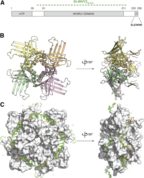
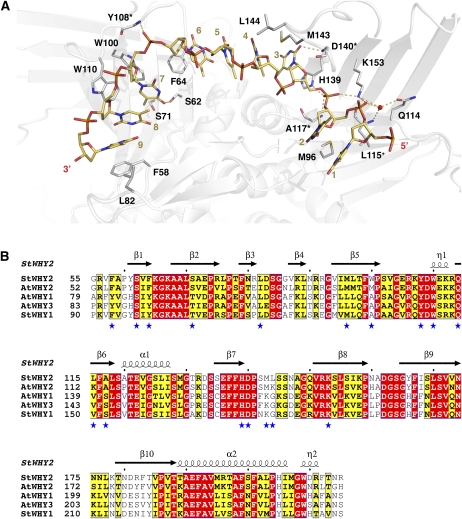
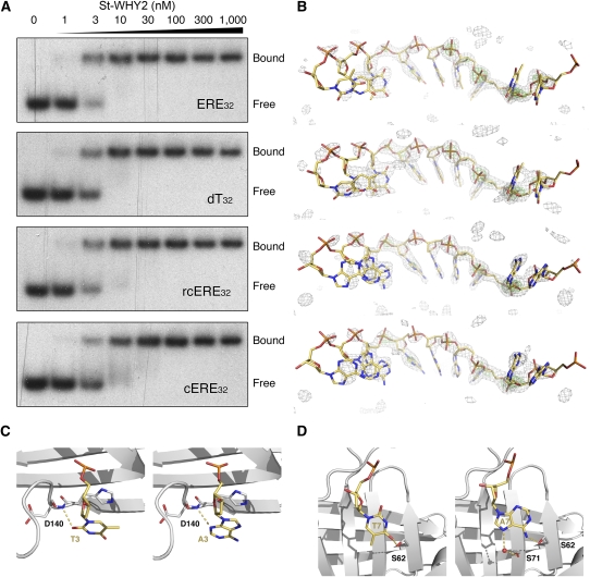
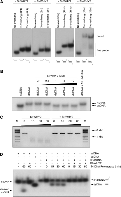
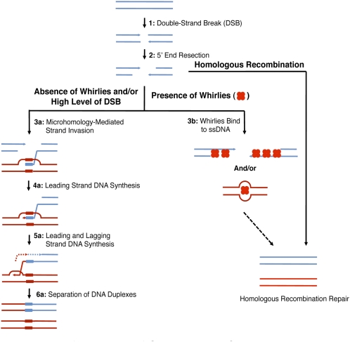
DNA double-strand breaks are highly detrimental to all organisms and need to be quickly and accurately repaired. Although several proteins are known to maintain plastid and mitochondrial genome stability in plants, little is known about the mechanisms of DNA repair in these organelles and the roles of specific proteins. Here, using ciprofloxacin as a DNA damaging agent specific to the organelles, we show that plastids and mitochondria can repair DNA double-strand breaks through an error-prone pathway similar to the microhomology-mediated break-induced replication observed in humans, yeast, and bacteria. This pathway is negatively regulated by the single-stranded DNA (ssDNA) binding proteins from the Whirly family, thus indicating that these proteins could contribute to the accurate repair of plant organelle genomes. To understand the role of Whirly proteins in this process, we solved the crystal structures of several Whirly-DNA complexes. These reveal a nonsequence-specific ssDNA binding mechanism in which DNA is stabilized between domains of adjacent subunits and rendered unavailable for duplex formation and/or protein interactions. Our results suggest a model in which the binding of Whirly proteins to ssDNA would favor accurate repair of DNA double-strand breaks over an error-prone microhomology-mediated break-induced replication repair pathway.


Figures

Figure 1.
Figure 1.
PCR Strategy to Detect DNA Rearrangements. Outward-facing PCR primers are used to monitor DNA duplication/circularization events, whereas inward-facing PCR primers detect deletions. In both cases, PCR amplification occurs only if a DNA rearrangement brings together the annealing sites of the primers. Gray areas represent repeated sequences.
Figure 2.
Figure 2.
DNA Rearrangements Accumulate in Plastids Following Treatment with the Gyrase Inhibitor Ciprofloxacin. (A) and (E) Phenotypic effects of various concentrations of ciprofloxacin (A) or novobiocin (E) on wild-type (WT) and why1 why3 Arabidopsis plants. Plants were grown for 3 weeks on solid media containing the indicated concentrations of ciprofloxacin (CIP) or novobiocin (NOV). (B) and (F) Histograms showing the average ± sd of plants with etiolated/variegated leaves at each ciprofloxacin (B) or novobiocin (F) concentration for wild-type and why1 why3 genotypes. At least three independent experiments were done. Plants with partially or fully white first true leaves were scored as etiolated/variegated. ND, etiolated/variegated leaves could not be counted at 0.75 μM ciprofloxacin and 100 μM novobiocin because they were very small or absent. (C) and (G) Electrophoretic analysis of representative PCR performed with 13 outward- or inward-facing plastid genome-directed PCR primers on total leaf DNA of wild-type and why1 why3 plants treated with ciprofloxacin (C) or with 10 outward- or inward-facing plastid genome-directed PCR primers on total leaf DNA of wild-type and why1 why3 plants treated with novobiocin (G). Low cycle amplification of the YCF2 plastid gene was used as a loading control. The oligonucleotides used for each PCR are indicated. Individual bands were cut from the gel, cloned, and sequenced. DNA rearrangements are listed in Supplemental Data Set 1 online. (D) and (H) Histograms showing the number of PCR products in wild-type and why1 why3 plants as a function of ciprofloxacin (D) or novobiocin (H) concentration.
Figure 3.
Figure 3.
DNA Rearrangements Accumulate in Mitochondria Following Treatment with the Gyrase Inhibitor Ciprofloxacin. (A) and (E) Phenotypic effects of various concentrations of ciprofloxacin (A) or novobiocin (E) on wild-type (WT) and why2-1 Arabidopsis plants. Plants were grown for 3 weeks on solid media containing the indicated concentrations of ciprofloxacin (CIP) or novobiocin (NOV). (B) and (F) Histograms showing the average ± sd of plants with etiolated/variegated leaves at each ciprofloxacin (B) or novobiocin (F) concentration for wild-type and why2-1 genotypes. At least three independent experiments were done. Plants with partially or fully white first true leaves were scored as etiolated/variegated. ND, etiolated/variegated leaves could not be counted at 0.75 μM ciprofloxacin and 100 μM novobiocin because they were very small or absent. (C) and (G) Electrophoretic analysis of representative PCR performed with 20 outward- or inward-facing mitochondrial genome-directed PCR primers on total leaf DNA of wild-type and why2-1 plants treated with ciprofloxacin (C) or with 10 outward- or inward-facing mitochondrial genome-directed PCR primers on total leaf DNA of wild-type and why2-1 plants treated with novobiocin (G). Low cycle amplification of the COX1 mitochondrial gene was used as a loading control. The oligonucleotides used for each PCR are indicated. Individual bands were cut from the gel, cloned, and sequenced. DNA rearrangements are listed in Supplemental Data Set 1 online. (D) and (H) Histograms showing the number of PCR products in wild-type and why2-1 plants as a function of ciprofloxacin (D) or novobiocin (H) concentration.
Figure 4.
Figure 4.
Crystal Structures of St-WHY2 in the Free Form and Bound to ERE32 at 2.2-Å Resolution. (A) Schematic representation of St-WHY2. Filled boxes indicate the position of the mitochondria transit peptide (mTP), the Whirly domain, and the acidic/aromatic C-terminal tail (CT). A green dotted line represents the construct used for structure determination. (B) Overall view of St-WHY2 in the free form in cartoon representation. The tetramer was generated by applying crystallographic fourfold symmetry. Protomers are colored in yellow, orange, pink, and green. (C) Surface representation of the protein moiety in the St-WHY2-ERE32 complex. The tetramer was generated by applying the crystallographic symmetry along the fourfold axis. Difference electron density was calculated from a Fo-Fc simulated annealing omit map encompassing DNA, contoured at 3.0 σ, and colored in green. The density was carved at 8 Å around the protein model.
Figure 5.
Figure 5.
Mechanism of ssDNA Binding of Whirly Proteins. (A) Protein–DNA interactions in the St-WHY2-ERE32 complex. The DNA and the DNA-interacting residues are in stick representation with carbon atoms colored in yellow and in gray, respectively. Nucleotides and protein residues are labeled. Asterisks, residues that contact DNA through their main-chain. +, residues that contact the DNA through both its main chain and side chain. Hydrogen bonds are represented as yellow dashed lines. A water molecule is represented as a red sphere. (B) Sequence alignment of the Whirly domain of Whirly proteins from Arabidopsis (At-WHY1, At-WHY2, and At-WHY3) and S. tuberosum (St-WHY1 and St-WHY2) with schematic secondary structural elements from St-WHY2. Secondary structure conformations are denoted at the top of the sequence alignment. α, α-helix; β, β-strand; η, 310 helix. Similarity above 70% is depicted in yellow, whereas perfect conservation is depicted in red. Blue stars underneath the sequence alignment indicate residues of St-WHY2 that interact with ssDNA. The alignments were made using T-coffee (Notredame et al., 2000) and the figure prepared using ESPript (Gouet et al., 2003).
Figure 6.
Figure 6.
WHY2 Binds ssDNA with Limited Sequence Specificity. (A) Representative EMSA results showing the binding of St-WHY2 to four different ssDNA sequences. Increasing amounts of WHY2 were incubated with target oligonucleotides ERE32, dT32, rcERE32, or cERE32 and the complexes resolved on a 10% (w/v) polyacrylamide gel. The sequences of these oligonucleotides can be found in Table 3. (B) Crystal structures of four different ssDNA sequences bound by St-WHY2. DNA molecules are presented as stick models with carbon atoms colored in yellow. Fo-Fc simulated annealing omit maps encompassing the entire DNA are contoured at 2.5 σ (colored in gray) or at 5 σ (colored in green). DNA molecules are presented in the same order as in (A). (C) and (D) Interactions between St-WHY2 and the edges of T3 (left panel) and A3 (right panel) (C) or the edges of T7 (left panel) and A7 (right panel) (D) in the WHY2-ERE32 and WHY2-rcERE32 structures, respectively. The representation and the orientation of the molecule are similar, thus revealing that compensating interactions enable WHY2 to bind DNA nucleobases that differ in size and in functional groups at these positions. A red sphere corresponds to a water molecule.
Figure 7.
Figure 7.
WHY2 Binds Single-Stranded Overhangs, Destabilizes dsDNA, and Protects ssDNA against Nuclease Degradation. (A) Representative EMSA results showing the binding of St-WHY2 to DNA duplexes with or without single-stranded dT8 or dT16 overhangs. Fifty nanomolar of WHY2 were incubated with target radiolabeled oligonucleotides and the complexes resolved on a 10% (w/v) polyacrylamide gel. Diagrams at the bottom of the gels illustrate the DNA used in the assay. An asterisk indicates the strand that is radiolabeled. (B) WHY2 destabilizes a DNA duplex. A DNA duplex with one strand radiolabeled was incubated with increasing amounts of St-WHY2 or BSA. After protein denaturation, DNA was resolved on a 7.5% acrylamide gel. (C) WHY2 protects DNA against mung bean nuclease degradation. Phage M13mp18 ssDNA either alone or prebound with St-WHY2 at a 1:10 protein/nucleotide ratio was incubated with mung bean nuclease for the indicated amount of time. After protein denaturation, DNA was resolved on an agarose gel. Black/white inverted images are shown. M represents the molecular weight markers. (D) WHY2 protects DNA against the exonuclease activity of T4 DNA Polymerase. Radiolabeled ssDNA or dsDNA with a 16-nucleotide 3′-overhang was complexed with St-WHY2 and then incubated with T4 DNA polymerase for the indicated amount of time. After protein denaturation, DNA was resolved on a 10% acrylamide gel. ssDNA, YG16_3; 3′-dsDNA, YG16_3-YC duplex; dsDNA, YG-YC duplex.
Figure 8.
Figure 8.
Model for the Repair of Organellar Double-Strand Breaks in the Absence or Presence of Whirly Proteins. Upon formation of a DSB (1), the 5′ end of the broken DNA molecule is resected from the break, exposing a 3′ tail (2). At this step, the break can be repaired through homologous recombination in a Whirly-independent manner. Alternatively, if the Whirlies are absent or the repair machinery is overloaded due to numerous DSBs, the 3′ tail can anneal to any exposed ssDNA through microhomologies (3a). A D-loop forms and DNA polymerization proceeds from the microhomology junction (4a). A replication fork is established and lagging strand synthesis initiates while leading strand synthesis continues (5a). DNA synthesis continues until the end of the chromosome is reached (6a). Alternatively, if the Whirlies are present and the DSB level is low (3b), Whirlies could bind and protect ssDNA (either the 3′ tail and/or any exposed ssDNA), thereby promoting homologous recombination and accurate DNA repair. Arrowheads represent 3′ ends; a box symbolizes the microhomology between broken and unbroken DNA molecules; dashed arrows in tandem represent lagging strand synthesis. [See online article for color version of this figure.]

References

    1. Abdelnoor R.V., Yule R., Elo A., Christensen A.C., Meyer-Gauen G., Mackenzie S.A. (2003). Substoichiometric shifting in the plant mitochondrial genome is influenced by a gene homologous to MutS. Proc. Natl. Acad. Sci. USA 100: 5968–5973 - PMC - PubMed
    1. Adams P.D., Grosse-Kunstleve R.W., Hung L.W., Ioerger T.R., McCoy A.J., Moriarty N.W., Read R.J., Sacchettini J.C., Sauter N.K., Terwilliger T.C. (2002). PHENIX: Building new software for automated crystallographic structure determination. Acta Crystallogr. D Biol. Crystallogr. 58: 1948–1954 - PubMed
    1. André C., Levy A.A., Walbot V. (1992). Small repeated sequences and the structure of plant mitochondrial genomes. Trends Genet. 8: 128–132 - PubMed
    1. Arrieta-Montiel M.P., Shedge V., Davila J., Christensen A.C., Mackenzie S.A. (2009). Diversity of the Arabidopsis mitochondrial genome occurs via nuclear-controlled recombination activity. Genetics 183: 1261–1268 - PMC - PubMed
    1. Baugnet-Mahieu L., Goutier R., Baes C. (1971). Differential response of mitochondrial and nuclear DNA syntheses to hydroxyurea in normal and regenerating rat liver. Biochem. Pharmacol. 20: 141–149 - PubMed

Publication types