. 2010 Oct 14;116(15):e66-73.
doi: 10.1182/blood-2010-04-278192. Epub 2010 Jun 15.

Discovery of novel mechanosensitive genes in vivo using mouse carotid artery endothelium exposed to disturbed flow

Affiliations

Discovery of novel mechanosensitive genes in vivo using mouse carotid artery endothelium exposed to disturbed flow

Chih-Wen Ni et al. Blood. .

Abstract

Recently, we showed that disturbed flow caused by a partial ligation of mouse carotid artery rapidly induces atherosclerosis. Here, we identified mechanosensitive genes in vivo through a genome-wide microarray study using mouse endothelial RNAs isolated from the flow-disturbed left and the undisturbed right common carotid artery. We found 62 and 523 genes that changed significantly by 12 hours and 48 hours after ligation, respectively. The results were validated by quantitative polymerase chain reaction for 44 of 46 tested genes. This array study discovered numerous novel mechanosensitive genes, including Lmo4, klk10, and dhh, while confirming well-known ones, such as Klf2, eNOS, and BMP4. Four genes were further validated for protein, including LMO4, which showed higher expression in mouse aortic arch and in human coronary endothelium in an asymmetric pattern. Comparison of in vivo, ex vivo, and in vitro endothelial gene expression profiles indicates that numerous in vivo mechanosensitive genes appear to be lost or dysregulated during culture. Gene ontology analyses show that disturbed flow regulates genes involved in cell proliferation and morphology by 12 hours, followed by inflammatory and immune responses by 48 hours. Determining the functional importance of these novel mechanosensitive genes may provide important insights into understanding vascular biology and atherosclerosis.


Figures

Figure 1
Figure 1
Global gene expression profiles in response to disturbed flow in mouse carotid artery endothelium in vivo. Total RNAs were obtained from intima of mouse left carotid (flow-disturbed, LCA) and right carotid (contralateral control, RCA) 12 and 48 hours after ligation. Illumina BeadChips containing 45 281 mouse genome-wide probes were used for the array study. Scatter plots show normalized intensities of each probe under 2 experimental conditions: LCA versus RCA at 12 hours (A) and 48 hours (B) after ligation. Genes that were up-regulated (red) or down-regulated (blue) (≥ 1.5-fold) at the FDR (≤ 10%) in LCA compared with RCA. (C-D) Hierarchical clustering analyses of mechanosensitive genes found in LCA endothelium compared with that of RCA are shown as heat maps. Each column represents a single sample pooled from 3 different LCAs or RCAs, and each row represents a single gene probe. (E) Venn diagrams show the temporal effects of disturbed flow on the number of up- or down-regulated mechanosensitive genes.
Figure 2
Figure 2
Validation of mechanosensitive genes by quantitative PCR. Total RNAs from intima of LCA or RCA at different time points (12, 24, and 48 hours) after ligation were collected. Differentially expressed genes were selected for quantitative PCR analyses based on 48-hour microarray results. Each RNA sample at each time was pooled from 3 different mouse carotids, representing a total of 9 (n = 3) to 15 (n = 5) mice. Microarray results for 12- and 48-hour time points are shown as fold increase (A) or fold decrease (C) of genes expressed in LCA over RCA in log2 scale (mean ± SEM; n = 3). All genes shown in the graphs were significant at FDR less than 10% at 48 hours. ‡Genes were also statistically significant (FDR < 10%) at 12 hours. Quantitative PCR validation results for 12-, 24-, and 48-hour time points are shown as fold increase (B) or fold decrease (D) of genes expressed in LCA over RCA in log2 scale (mean ± SEM; n = 3-5). All genes shown in the graphs were significant (P < .05) at 48 hours. *Genes were also statistically significant (P < .05) at 12 and 48 hours. (E) Six genes of interests that did not reach statistical significance (> 10% FDR) were examined by quantitative PCR. Data are mean ± SEM (n = 3). ‡< 10% FDR (LCA vs RCA). * < .05 (LCA vs RCA).
Figure 3
Figure 3
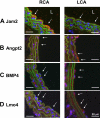
Disturbed flow in LCA decreases protein expression of Jam2 while up-regulating Angpt2, BMP4, and Lmo4. C57BL/6 mice underwent partial ligation, and LCA and RCA were collected 2 days after ligation. Paraffin sections were stained with specific antibodies for Jam2 (A), Angpt2 (B), BMP4 (C), and Lmo4 (D). Nuclei were counterstained with Hoechst (blue). Arrows indicate the protein expression in endothelial cells. L indicates lumen. Images are representative of n = 4 mice.
Figure 4
Figure 4
Lmo4 is differentially expressed in mouse aortic arch and human coronary artery. (A) En face staining of greater curvature (GC), lesser curvature (LC) of the arch, and the thoracic aorta (TA) was performed with Lmo4 antibody (red). Blue signal indicates nuclei stained with 4,6-diamidino-2-phenylindole; green signal indicates elastic laminae detected by autofluorescence. Shown are representative images of 7 different mice. (B) Paraffin sections of human left anterior descending coronary artery were stained for Lmo4 protein expression. Overall staining patterns were shown at low magnification (original magnification ×5) as a composite figure and zoomed views (original magnification ×20) of the indicated areas (broken box). The raw images of the composite figure are shown in supplemental Figure 3.

Comment in

Similar articles

Cited by

References

    1. Ross R. Atherosclerosis: an inflammatory disease. N Engl J Med. 1999;340(2):115–126. - PubMed
    1. Libby P. Inflammation in atherosclerosis. Nature. 2002;420(6917):868–874. - PubMed
    1. Ku DN, Giddens DP, Zarins CK, Glagov S. Pulsatile flow and atherosclerosis in the human carotid bifurcation: positive correlation between plaque location and low oscillating shear stress. Arteriosclerosis. 1985;5(3):293–302. - PubMed
    1. VanderLaan PA, Reardon CA, Getz GS. Site specificity of atherosclerosis: site-selective responses to atherosclerotic modulators. Arterioscler Thromb Vasc Biol. 2004;24(1):12–22. - PubMed
    1. Berk BC. Atheroprotective signaling mechanisms activated by steady laminar flow in endothelial cells. Circulation. 2008;117(8):1082–1089. - PubMed

Publication types