. 2010 Aug 27;285(35):27057-27066.
doi: 10.1074/jbc.M110.112276. Epub 2010 Jun 15.

FEN1 ensures telomere stability by facilitating replication fork re-initiation

Affiliations

FEN1 ensures telomere stability by facilitating replication fork re-initiation

Abhishek Saharia et al. J Biol Chem. .

Abstract

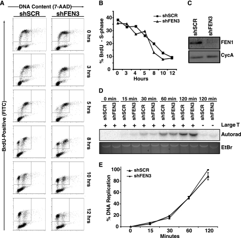
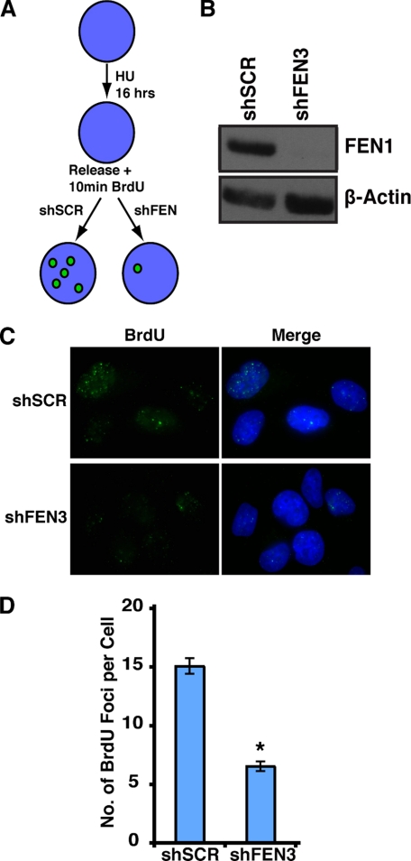
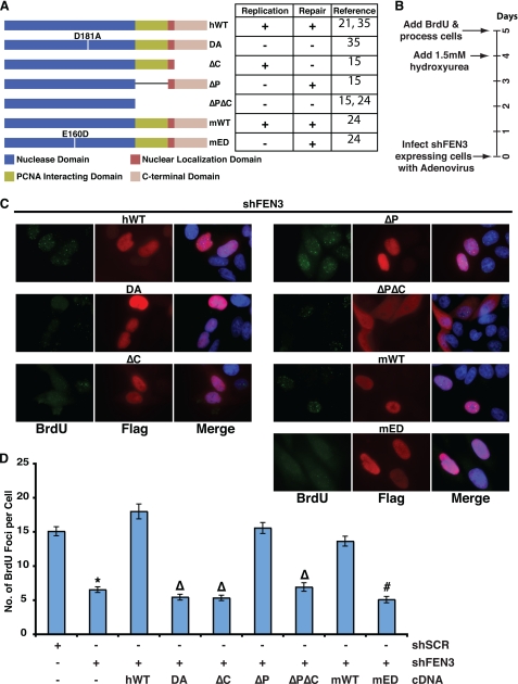
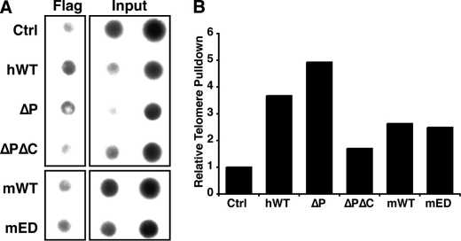
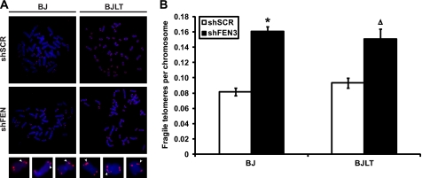
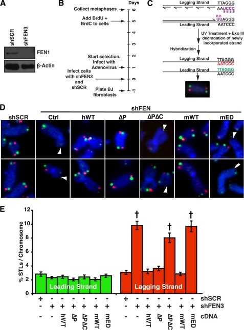
Telomeres are terminal repetitive DNA sequences whose stability requires the coordinated actions of telomere-binding proteins and the DNA replication and repair machinery. Recently, we demonstrated that the DNA replication and repair protein Flap endonuclease 1 (FEN1) is required for replication of lagging strand telomeres. Here, we demonstrate for the first time that FEN1 is required for efficient re-initiation of stalled replication forks. At the telomere, we find that FEN1 depletion results in replicative stress as evidenced by fragile telomere expression and sister telomere loss. We show that FEN1 participation in Okazaki fragment processing is not required for efficient telomere replication. Instead we find that FEN1 gap endonuclease activity, which processes DNA structures resembling stalled replication forks, and the FEN1 interaction with the RecQ helicases are vital for telomere stability. Finally, we find that FEN1 depletion neither impacts cell cycle progression nor in vitro DNA replication through non-telomeric sequences. Our finding that FEN1 is required for efficient replication fork re-initiation strongly suggests that the fragile telomere expression and sister telomere losses observed upon FEN1 depletion are the direct result of replication fork collapse. Together, these findings suggest that other nucleases compensate for FEN1 loss throughout the genome during DNA replication but fail to do so at the telomere. We propose that FEN1 maintains stable telomeres by facilitating replication through the G-rich lagging strand telomere, thereby ensuring high fidelity telomere replication.


Figures

FIGURE 1.
FIGURE 1.
FEN1 depletion does not affect S phase progression or in vitro DNA replication. A, cell cycle progression of HeLa cells expressing shSCR or shFEN3 is shown. HeLa cells were labeled with BrdU for 1 h and analyzed at the indicated times using an anti-BrdU antibody (FITC-conjugated) and 7-amino-actinomysin D (7-AAD) to label DNA content. BrdU-positive cells are displayed on the y axis and represent cells that transit through S phase during BrdU labeling. The x axis displays the DNA content of the cells as indicated by incorporation of 7-amino-actinomycin D (G1 and G2/M cells have a 2 n and 4 n content of DNA, respectively). B, quantification of the percent of BrdU-positive cells in S phase after BrdU pulse (representative experiment is shown) is shown. The cells present in the inset boxes in A are BrdU-positive and consist of cells in G1, S, and G2/M phases of the cell cycle. Only the S phase cells (those that are present between G1 and G2 (2n and 4n DNA content, respectively) within the BrdU-positive population are plotted on the graph. Error bars represent S.E. C, shown are Western blots of S100 lysates from control and FEN1-depleted HeLa cells. Cyclophilin A (CycA, lower panel) is shown as a loading control. D, an SV40 Large T antigen-dependent in vitro DNA replication assay was conducted using lysates from control (shSCR) and FEN1-depleted (shFEN3) HeLa cells as described under “Experimental Procedures.” The assay was stopped at the indicated times, and the replication products were separated via gel electrophoresis. The replication products were detected via autoradiography (Autorad), and the input DNA was observed via ethidium bromide (EtBr) staining. E, shown is quantification of the replication products at the indicated times in D. Two independent experiments were conducted in duplicate, and the average of the four experiments is shown. The error bars represent S.E.
FIGURE 2.
FIGURE 2.
FEN1 depletion decreases re-initiation of stalled replication forks. A, shown is a schematic of the stalled replication fork re-initiation assay and the expected results. HU, hydroxyurea. B, a Western blot analysis shows FEN1 depletion. Short hairpins against FEN1 (shFEN3) or a scrambled sequence (shSCR) were expressed in HeLa cells. FEN1 (upper panel) and β-actin (lower panel) protein levels were assessed by Western blot analysis. C, representative images show that FEN1 depletion decreases BrdU incorporation in hydroxyurea-treated cells. Immunofluorescence was conducted using an anti-BrdU antibody (green) and 4′,6-diamidino-2-phenylindole (DAPI, blue). D, quantification of the number of BrdU foci per cell in HeLa cells transduced with the indicated shRNA is shown. BrdU foci in no fewer than 100 cells were counted for each condition, and the experiment was conducted twice (a representative experiment is presented). Error bars represent S.E. (*, p < 0.0001).
FIGURE 3.
FIGURE 3.
The gap endonuclease activity and C terminus of FEN1 are essential to re-initiate stalled replication forks. A, the schematic shows the different FEN1 alleles used in the study. Inferences on whether the different FEN1 alleles are replication competent or repair competent are shown on the right of the schematic with their associated references. These inferences were made based on nuclease activity and ability to interact with the WRN and PCNA proteins. The mutant proteins are as follows: ΔC (amino acids 360–380 deleted), ΔP (amino acids 337–344 deleted), and ΔPΔC (amino acids 337–380 deleted). B, the timeline of the experimental procedure is given in days. C, representative images show BrdU incorporation after hydroxyurea treatment in FEN1-depleted cells expressing wild-type or FEN1 mutants. Immunofluorescence was conducted using an anti-BrdU (green) antibody, anti-FLAG (red) antibody, and DAPI (blue). D, quantification of the number of BrdU foci per cell in FEN1-depleted HeLa cells with the indicated ectopic FEN1 expression (wild-type or mutant) is shown. Only cells expressing FLAG-tagged FEN1 (marked by red in C) was quantified. No fewer than 75 cells were counted for each condition, and the experiment was conducted twice (a representative experiment is presented). The error bars represent S.E. (*, p < 0.0001 compared with shSCR; Δ, p < 0.0001 compared with hWT; #, p < 0.0001 compared with mWT).
FIGURE 4.
FIGURE 4.
FEN1 mutants localize to the telomere. A, FEN1 alleles localize to the telomere. Representative ChIP analysis of 293T cells (Ctrl) or 293T cells transfected with wild-type FEN1 (hWT or mWT) or FEN1 mutants (ΔP, ΔPΔC, or mED), subjected to immunoprecipitation with the FLAG (M2) antibody. Precipitated DNA was probed for the presence of telomeric sequences as described under “Experimental Procedures.” The inputs indicate 0.1 and 0.2% of total. B, quantification of the representative ChIP assay is shown. Percent of telomeric DNA immunoprecipitated with the FLAG antibody was calculated using input DNA, and the control pulldown percentage was set to 1.
FIGURE 5.
FIGURE 5.
FEN1 depletion results in fragile site expression at telomeres. A, representative FISH of metaphases obtained from BJ fibroblasts (BJ) or BJ fibroblasts expressing SV40 Large T antigen and telomerase (BJLT). Cells expressing a control hairpin (shSCR) or depleted of FEN1 (shFEN3) are indicated. Chromosomes were hybridized with the PNA telomere probe Cy3-(CCCTAA)3 (red) and stained with DAPI (blue). Magnified images show representative fragile telomeres (arrowheads). B, quantification of telomere fragility, depicted as the number of fragile telomeres observed per chromosome. No fewer than 60 metaphases from two independent experiments were analyzed for each condition, and an average of the two experiments is shown (*, p < 0.0001; Δ, p < 0.001). Error bars represent S.E.
FIGURE 6.
FIGURE 6.
The gap endonuclease activity of FEN1 is essential for its function at the telomere. A, Western blot analysis of FEN1 (upper panel) from BJ fibroblasts expressing a control hairpin (shSCR) or depleted of FEN1 (shFEN3) is shown. β-Actin (lower panel) is shown as a loading control. B, shown is a timeline of experimental procedure given in days. C, a CO-FISH schematic is shown. Newly synthesized DNA strands incorporate BrdU and BrdC. UV and ExoIII treatment resulted in degradation of newly synthesized DNA containing BrdU and BrdC, and the template strands were hybridized with Cy3-(CCCTAA)3 (red, lagging strand) and fluorescein-(TTAGGG)3 (green, leading strand) PNA probes (25). D, representative CO-FISH of chromosomes from BJ fibroblasts expressing shSCR or shFEN3 and the indicated FEN1 alleles is shown. Ctrl refers to cells that do not express exogenous FEN1 protein. Color schemes are as described in C. DNA was stained with DAPI (blue). The arrowheads indicate missing telomeres. E, shown is quantification of STLs on metaphase chromosomes after depletion of endogenous FEN1 and expression of the indicated FEN1 allele, depicted as percentage of chromosomes with missing leading (green) and lagging (red) strand telomeres. A minimum of 60 metaphases from two independent experiments was analyzed per treatment in a blinded fashion, and an average of the two experiments is shown (*, p < 0.0001). The error bars represent S.E.

Similar articles

Cited by

References

    1. Wu L., Hickson I. D. (2002) Mutat. Res. 509, 35–47 - PubMed
    1. Sidorova J. M. (2008) DNA Repair 7, 1776–1786 - PMC - PubMed
    1. Singh D. K., Ahn B., Bohr V. A. (2009) Biogerontology 10, 235–252 - PMC - PubMed
    1. Gilson E., Géli V. (2007) Nat. Rev. Mol. Cell Biol. 8, 825–838 - PubMed
    1. Verdun R. E., Karlseder J. (2007) Nature 447, 924–931 - PubMed

Publication types