. 2010 Sep;18(9):1706-13.
doi: 10.1038/mt.2010.100. Epub 2010 Jun 15.

Macrophages expressing heme oxygenase-1 improve renal function in ischemia/reperfusion injury

Affiliations

Macrophages expressing heme oxygenase-1 improve renal function in ischemia/reperfusion injury

David A Ferenbach et al. Mol Ther. 2010 Sep.

Abstract

Acute kidney injury has a high mortality and lacks specific therapies, with ischemia/reperfusion injury (IRI) being the predominant cause. Macrophages (M phi) have been used successfully in cell therapy to deliver targeted therapeutic genes in models of inflammatory kidney disease. Heme oxygenase-1 (HO-1) catalyzes heme breakdown and has important cytoprotective functions. We hypothesized that administration of M phi modified to overexpress HO-1 would protect from renal IRI. Using an adenoviral construct (Ad-HO-1), HO-1 was overexpressed in primary bone marrow-derived M phi (BMDM). In vitro Ad-HO-1 M phi showed an anti-inflammatory phenotype with increased phagocytosis of apoptotic cells (ACs) and increased interleukin (IL)-10 but reduced TNF-alpha and nitric oxide (NO) following lipopolysaccharide/interferon-gamma (IFN gamma) stimulation compared to control transduced or unmodified M phi. In vivo, intravenously (IV) injected M phi homed preferentially to the post-IRI kidney compared to uninjured control following experimental IRI. At 24 hours postinjury, despite equivalent levels of tubular necrosis, apoptosis, and capillary density between groups, the injection of Ad-HO-1 M phi resulted in preserved renal function (serum creatinine reduced by 46%), and reduced microvascular platelet deposition. These data demonstrate that genetically modified M phi improve the outcomes in IRI when administered after the establishment of structural injury, raising the prospect of targeted cell therapy to support the function of the acutely injured kidney.


Figures

Figure 1
Figure 1
Adenoviral transduction results in expression of HO-1 protein in Mφ. (a) Western blotting demonstrates potent induction of HO-1 protein expression in BMDM following increasing multiplicity of infection with Ad-HO-1. (b) HO-1 transduction was associated with increased bioactivity as shown by degradation of heme to bilirubin (n = 8/group). BMDM, bone marrow–derived Mφ.
Figure 2
Figure 2
Transduction of HO-1 in Mφ results in altered responses to classical activating stimuli. Transduction with Ad-HO-1 results in significantly reduced (a) Mφ NO and (b) TNFα production, with augmented (c) IL-10 in response to stimulation with IFNγ+LPS (n = 9/group; all P < 0.001 versus all groups by analysis of variance).
Figure 3
Figure 3
Transduction of HO-1 in Mφ results in increased phagocytosis of apoptotic cells in vitro. Transduction with Ad-HO-1 results in (a) augmented phagocytosis of apoptotic cells (ACs) (n = 9/group; P = 0.0032) and (b) increased phagocytic index (n = 9/group; P = 0.0093). Phagocytosis was quantified visually using colocalization of Lysotracker Red–labeled Mφ and CM-Green-labeled apoptotic cells to confirm presence of the apoptotic cells within a phagolysosome. Photomicrographs of Mφ following a 30-minute interaction with apoptotic cells [(c) control unmodified Mφ, (d) Ad-HO-1 Mφ phagocytosed ACs shown with white arrows]. BMDM, bone marrow–derived Mφ.
Figure 4
Figure 4
Intravenously administered Mφ home to the injured kidney after IRI. (a) IV administered Mφ localize selectively to the injured kidney compared to the contralateral uninjured control (P = 0.0048, n = 6/group). (b) Maximal localization is seen at 1 hour after injection. PKH+ Mφ can be visualized within the interstitium of the injured renal medulla at (c) 1 and (d) 24 hours (original magnification: ×200, blue: DAPI nuclear stain, green: tissue autofluorescence, red: PKH26+ Mφ). Medullary F4/80+ cell counts (mean per ×400 field) are augmented in animals receiving IV Mφ. (e) Exogenously delivered Mφ were identified by PKH+ fluorescence, and comprise between 35 and 58% of total medullary Mφ post-IRI. IRI, ischemia/reperfusion injury.
Figure 5
Figure 5
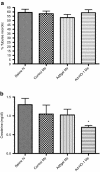
Treatment with Ad-HO-1 transduced Mφ results in functional protection after IRI at comparable levels of acute tubular necrosis. (a) Cell therapy with Ad-HO-1 transduced Mφ after experimental IRI has no impact on severity of tubular necrosis but (b) results in significant preservation of renal function 24 hours after injury (all groups n = 9–12; P < 0.05). IRI, ischemia/reperfusion injury.
Figure 6
Figure 6
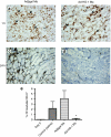
Animals treated with Ad-HO-1 transduced Mφ exhibit increased clearance of renal platelet deposition. Treatment with HO-1 expressing Mφ has no effect on immediate levels of platelet deposition compared with control Mφ at 1 hour post-IRI, while resulting in significant reduction in platelet deposition at 24 hours post-IRI [(a) Adβgal Mφ at 1 hour; (b) Ad-HO-1 Mφ at 1 hour, (c) Adβgal Mφ at 24 hours; (d) Ad-HO-1 Mφ at 24 hours] demonstrated in frozen sections stained with CD41 mAb (original magnification: ×200). (e) Mean area of platelet staining was quantified by image analysis and expressed as % hpf positive for CD41. HO-1, heme oxygenase-1; IRI, ischemia/reperfusion injury.
Figure 7
Figure 7
Proposed schema of renal dysfunction in the aftermath of IRI. Dotted arrows indicate pathways potentially ameliorated by actions of HO-1 expressing Mφ. IRI, ischemia/reperfusion injury.

Similar articles

Cited by

References

    1. Thadhani R, Pascual M., and, Bonventre JV. Acute renal failure. N Engl J Med. 1996;334:1448–1460. - PubMed
    1. Bonventre JV., and, Zuk A. Ischemic acute renal failure: an inflammatory disease. Kidney Int. 2004;66:480–485. - PubMed
    1. Day YJ, Huang L, Ye H, Linden J., and, Okusa MD. Renal ischemia-reperfusion injury and adenosine 2A receptor-mediated tissue protection: role of macrophages. Am J Physiol Renal Physiol. 2005;288:F722–F731. - PubMed
    1. Miura M, Fu X, Zhang QW, Remick DG., and, Fairchild RL. Neutralization of Gro alpha and macrophage inflammatory protein-2 attenuates renal ischemia/reperfusion injury. Am J Pathol. 2001;159:2137–2145. - PMC - PubMed
    1. Burne-Taney MJ, Ascon DB, Daniels F, Racusen L, Baldwin W., and, Rabb H. B cell deficiency confers protection from renal ischemia reperfusion injury. J Immunol. 2003;171:3210–3215. - PubMed

Publication types

MeSH terms

LinkOut - more resources