. 2010 Jun 11;5(6):e11025.
doi: 10.1371/journal.pone.0011025.

Host factors required for modulation of phagosome biogenesis and proliferation of Francisella tularensis within the cytosol

Affiliations

Host factors required for modulation of phagosome biogenesis and proliferation of Francisella tularensis within the cytosol

Christine Akimana et al. PLoS One. .

Abstract

Francisella tularensis is a highly infectious facultative intracellular bacterium that can be transmitted between mammals by arthropod vectors. Similar to many other intracellular bacteria that replicate within the cytosol, such as Listeria, Shigella, Burkholderia, and Rickettsia, the virulence of F. tularensis depends on its ability to modulate biogenesis of its phagosome and to escape into the host cell cytosol where it proliferates. Recent studies have identified the F. tularensis genes required for modulation of phagosome biogenesis and escape into the host cell cytosol within human and arthropod-derived cells. However, the arthropod and mammalian host factors required for intracellular proliferation of F. tularensis are not known. We have utilized a forward genetic approach employing genome-wide RNAi screen in Drosophila melanogaster-derived cells. Screening a library of approximately 21,300 RNAi, we have identified at least 186 host factors required for intracellular bacterial proliferation. We silenced twelve mammalian homologues by RNAi in HEK293T cells and identified three conserved factors, the PI4 kinase PI4KCA, the ubiquitin hydrolase USP22, and the ubiquitin ligase CDC27, which are also required for replication in human cells. The PI4KCA and USP22 mammalian factors are not required for modulation of phagosome biogenesis or phagosomal escape but are required for proliferation within the cytosol. In contrast, the CDC27 ubiquitin ligase is required for evading lysosomal fusion and for phagosomal escape into the cytosol. Although F. tularensis interacts with the autophagy pathway during late stages of proliferation in mouse macrophages, this does not occur in human cells. Our data suggest that F. tularensis utilizes host ubiquitin turnover in distinct mechanisms during the phagosomal and cytosolic phases and phosphoinositide metabolism is essential for cytosolic proliferation of F. tularensis. Our data will facilitate deciphering molecular ecology, patho-adaptation of F. tularensis to the arthropod vector and its role in bacterial ecology and patho-evolution to infect mammals.


Conflict of interest statement

Competing Interests: The authors have declared that no competing interests exist.

Figures

Figure 1
Figure 1. D. melanogaster RNAi screen and identification of host factors required for proliferation of F. tularensis.
(A) Genome-wide screen design. Procedural outline for the screen including RNAi treatment, infection, and identification of hits from a z-score heat map. (B) Functional categories of RNAi targets that decreased intracellular proliferation of F. tularensis in the secondary screen and (C) functional categories of targets that were also identified to be required for intracellular proliferation of L. monocytogenes, Mycobacterium, or Chlamydia in the secondary screen. The number of host factors within each functional category in panels B and C are shown.
Figure 2
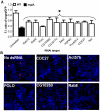
Figure 2. Representative host factors required for intracellular growth of F. tularensis.
S2R+ cells were treated with RNAi targeting gfp, an arbitrary negative control, or the indicated representative RNAi and infected with F. tularensis expressing GFP. (A) At 4 days post-infection, the fluorescence intensity (F.I.) of F. tularensis GFP was measured. The GFP-expressing mglA mutant that is severely defective in intracellular proliferation was used as a control. Results of representative genes were obtained in two independent experiments, each with up to 4 wells. Asterisk indicates RNAi that produced a statistically significant decrease in F. tularensis GFP fluorescence intensity versus control RNAi, as described in materials and methods. Error bars represent standard error of the mean. (B) Representative RNAi-treated cells were fixed and stained with Hoechst dye (blue) and bacteria were labeled with antibodies (red) at 24 h post-infection.
Figure 3
Figure 3. Suppression of intracellular replication of F. tularensis within CDC27 and USP22 RNAi-silenced HEK293T cells.
A. Representative microscopy images of HEK293T cells at 8 h after infection by F. tularensis. HEK293T cells were either left untreated or treated with the RNAi negative control or the corresponding RNAi for USP22 or CDC27 for 48 h before infection, then infected with MOI of 10 by the WT strain U112 for 1 hr, followed with 1 hr gentamicin and additional 6 h of incubation for a total of 8 h. B. Western blot results confirming specific gene silencing. Membranes were blotted with specific antibodies against USP22 or CDC27, then stripped and re-probed with the anti-actin antibody as a loading control. C. Effect of USP22 and CDC27 silencing on bacterial replication after 8 h of infection. HEK293T cells were either left untreated or RNAi treated for 48 h before infection, then infected with the WT strain U112. We considered cells harboring 6–15 bacteria after 8 h of infection as normal WT levels of replication. At least 100 infected cells from different field were analyzed in each experiment. Data are the results of one experiment representative of three independent experiments. The asterisk indicates statistically significant differences between the control and the USP22 and CDC27 RNAi- treated cells.
Figure 4
Figure 4. Late cessation of intracellular proliferation within PI4KCA-silenced cells.
A) Representative microscopy images of HEK293T cells after 8 and 24 h infection by F. tularensis. HEK293T cells were either treated with the RNAi negative control or the PI4KCA RNAi for 48 h before infection, then infected with MOI of 10 with the WT strain U112 for 1 hr, followed by 1 h gentamicin and an additional 6 h (left panel) or 22 h (right panel) of incubation. B. Western blot results confirming PI4KCA gene silencing. Membranes were blotted with anti-PI4KCA antibody, then stripped and re-probed with the anti-actin antibody as our loading control. C. Effect of PI4KCA silencing on bacterial replication after 24 h of infection. HEK293T cells were either left untreated or RNAi treated for 48 h before infection, then infected with the WT strain. Depending on the MOI that we used, we considered cells harboring 6–15 bacteria after 8 h of infection and more than 25 bacteria after 24 h to reflect the normal WT levels of replication. At least 100 infected cells from different fields were analyzed in each experiment. Data are the results of one experiment representative of three independent experiments. The asterisk indicates statistically significant difference between the control and the PI4KCA RNAi- treated cells.
Figure 5
Figure 5. CDC27 but not USP22 or PI4KCA is required for modulation of phagosome biogenesis.
A. Representative confocal microscopy images that show co-localization of the late endosomal/lysosomal marker LAMP-2 with the phagosomes harboring the iglC mutant in the untreated HEK293T cells (UN) as well as the WT strain in the RNAi-treated cells at 2 h after infection. B. Representative confocal microscopy images that show co-localization of the lysosomal enzyme cathepsin D with the phagosomes harboring the iglC mutant in the untreated cells (UN), as well as the WT strain in the RNAi-treated cells. C. Quantification of co-localization of the phagosomes with LAMP-2 (L-2) and cathepsin-D (C–D) markers at various time points. The results are based on examination of at least 100 bacteria at 2 h after infection analyzed by confocal microscopy in 2 independent experiments performed in triplicate and the error bars represent standard deviation.
Figure 6
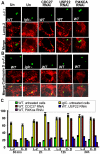
Figure 6. CDC27 but not USP22 or PI4KCA is required for bacterial escape into the cytosol.
Representative confocal microscopy images of the WT F. tularensis within untreated or RNAi-treated cells to determine phagosomal escape. Phagosomal escape was determined by the ability of GFP-expressing intracellular bacteria to bind anti-F. tularensis antibody (red) loaded into the host cell cytosol after preferential permeabilization of the plasma membrane, compared to the bacteria found within intact vacuoles (green) that are impermeable to the antibody. The % of cytosolic bacteria are shown, based on examination of at least 100 bacteria at 2 h after infection. The results are representative of three independent experiments performed in triplicate.

Similar articles

Cited by

References

    1. Santic M, Akimana C, Asare R, Kouokam JC, Atay S, et al. Intracellular fate of Francisella tularensis within arthropod-derived cells. Environ Microbiol. 2009;11:1473–1481. - PubMed
    1. Ellis J, Oyston PC, Green M, Titball RW. Tularemia. Clin Microbiol Rev. 2002;15:631–646. - PMC - PubMed
    1. Champion MD, Zeng Q, Nix EB, Nano FE, Keim P, et al. Comparative genomic characterization of Francisella tularensis strains belonging to low and high virulence subspecies. PLoS Pathog. 2009;5:e1000459. - PMC - PubMed
    1. Vogler AJ, Birdsell D, Price LB, Bowers JR, Beckstrom-Sternberg SM, et al. Phylogeography of Francisella tularensis: global expansion of a highly fit clone. J Bacteriol. 2009;191:2474–2484. - PMC - PubMed
    1. Titball R, Sjosted A. Francisella tularensis: an overview. ASM News. 2003;69:558–563.

Publication types

MeSH terms

Substances

LinkOut - more resources