. 2010 Jun 11;5(6):e11016.
doi: 10.1371/journal.pone.0011016.

Erythropoietin amplifies stroke-induced oligodendrogenesis in the rat

Affiliations

Erythropoietin amplifies stroke-induced oligodendrogenesis in the rat

Li Zhang et al. PLoS One. .

Abstract

Background: Erythropoietin (EPO), a hematopoietic cytokine, enhances neurogenesis and angiogenesis during stroke recovery. In the present study, we examined the effect of EPO on oligodendrogenesis in a rat model of embolic focal cerebral ischemia.

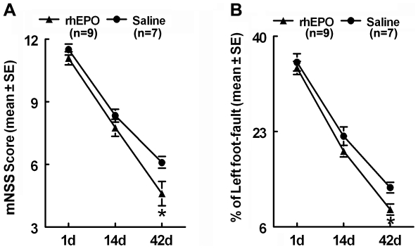
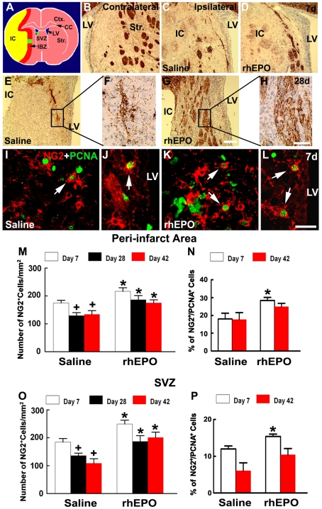
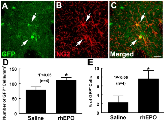
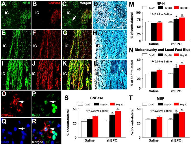
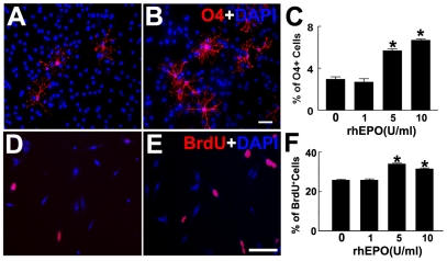
Methodology and principal findings: Recombinant human EPO (rhEPO) at a dose of 5,000 U/kg (n = 18) or saline (n = 18) was intraperitoneally administered daily for 7 days starting 24 h after stroke onset. Treatment with rhEPO augmented actively proliferating oligodendrocyte progenitor cells (OPCs) measured by NG2 immunoreactive cells within the peri-infarct white matter and the subventricular zone (SVZ), but did not protect against loss of myelinating oligodendrocytes measured by cyclic nucleotide phosphodiesterase (CNPase) positive cells 7 days after stroke. However, 28 and 42 days after stroke, treatment with rhEPO significantly increased myelinating oligodendrocytes and myelinated axons within the peri-infarct white matter. Using lentivirus to label subventricular zone (SVZ) neural progenitor cells, we found that in addition to the OPCs generated in the peri-infarct white matter, SVZ neural progenitor cells contributed to rhEPO-increased OPCs in the peri-infarct area. Using bromodeoxyuridine (BrdU) for birth-dating cells, we demonstrated that myelinating oligodendrocytes observed 28 days after stroke were derived from OPCs. Furthermore, rhEPO significantly improved neurological outcome 6 weeks after stroke. In vitro, rhEPO increased differentiation of adult SVZ neural progenitor cells into oligodendrocytes and enhanced immature oligodendrocyte cell proliferation.

Conclusions: Our in vivo and in vitro data indicate that EPO amplifies stroke-induced oligodendrogenesis that could facilitate axonal re-myelination and lead to functional recovery after stroke.


Conflict of interest statement

Competing Interests: The authors have declared that no competing interests exist.

Figures

Figure 1
Figure 1. Neurological functional outcome.
Panels A and B show the neurological functional outcome measured by mNSS (A) and foot-fault test (B) 1, 14, and 42 days after MCAo. Values are mean ± SE. *P<0.05 as compared with the saline-treated group.
Figure 2
Figure 2. Effects of EPO on oligodendrocytes.
Panel A is a schematic representation of a brain coronal section showing peri-infarct striatum (green box in A) and ipsilateral SVZ (green arrowhead in A) where all representative microscopic images were acquired. The yellow color represents ischemic core, and the red color indicates peri-infarct region. Panels B to H show CNPase immunoreactivity of representative rats treated with saline (B, C, E, F) and rhEPO (D, G, H), which were acquired from ipsilateral (C to H) and contralateral (B) striatum 7d (B to D) and 28d (E to H) after stroke. Panels F and H are high magnification images from boxed areas in panels E and G, respectively. Panels I to L show the double fluorescence immunostaining of NG2 (red) with PCNA (green), from representative rats treated with saline (I, J) and rhEPO (K, L), which were acquired from peri-infarct striatum (I, K) and SVZ (J, L). Panels M and O are the quantitative data of NG2 positive cells in peri-infarct corpus callosum and striatum (M), and ipsilateral SVZ (O). Panels N and P are the quantitative data of NG2 and PCNA double positive cells in peri-infarct corpus callosum and striatum (N), and ipsilateral SVZ (P). Values are mean ± SE. *P<0.05 vs saline. + P<0.05 vs the 7 day group. IBZ  =  ischemic boundary zone, CC  =  corpus callosum; Ctx  =  cortex; IC  =  ischemic core; LV  =  lateral ventricle; Str  =  striatum; SVZ  =  subventricular zone. Scale bars: 100 µm for panels B, C, D, E, and G, 50 µm for panels F and H. For 7 days, n = 4/saline and n = 4/rhEPO; for 28 days, n = 7/saline and n = 5/rhEPO; for 42 days, n =  7/saline and n = 9/rhEPO.
Figure 3
Figure 3. Effects of EPO on the recruitment of OPCs from the SVZ.
Panels A to C show GFP (green) and NG2 (red) immunoreactive cells in peri-infarct striatum from a representative rat treated with rhEPO 7 days after stroke. Some of GFP positive cells that were labeled in the SVZ 3 days prior to stroke were NG2 immunoreactive (arrows) in the peri-infarct area. Quantitative data show that the rhEPO treatment significantly increased number of GFP labeled cells (D) and percentage of NG2 positive GFP labeled cells (E) compared with the saline-treated group. *P<0.05 vs saline. Scale bars = 100 µm.
Figure 4
Figure 4. Effects of EPO on axons and myelinating oligodendrocytes.
Panels A to L show the double fluorescence immunostaining of NF-H (green, A, C, E, G, I, and K) with CNPase (red, B, C, F, G, J, and K), and Bielschowsky and Luxol fast blue staining (D, H, and L) from representative rats treated with saline (A to D, at 7d; E to H, at 42d) and rhEPO (I to L, at 42d), which were acquired from ipsilateral striatum. An arrow in panels A to C indicate NF-H positive axons with appearances of swellings and bulbs (green) that were not associated with CNPase immumoreactivity (red) 7d after stroke. Quantitative data show that rhEPO treatment significantly increase NF-H immunoreactivity (M) and Bielschowsky and Luxol fast blue (N) positive axons in the peri-infarct striatum compared with saline treated rats 28 and 42d after stroke. Confocal microscopic images (O to R) show that a CNPase immunoreactive cell (red, arrow) was BrdU positive (green, arrow) in the nucleus (DAPI, blue, arrow) of a representative rat treated with rhEPO 28d after stroke. Quantitative data shows that treatment with rhEPO significantly increases the CNPase (S) and MBP (T) immunoreactive area 28 and 42d after stroke. Values are mean ± SE. IC, ischemic core. Scale bars: 50 µm for panels A to L, 20 µm for panels E to H. IC  =  ischemic core. For 7 days, n = 4/saline and n = 4/rhEPO; for 28 days, n = 7/saline and n = 5/rhEPO; for 42 days, n = 7/saline and n = 9/rhEPO.
Figure 5
Figure 5. Effects of EPO on generation of OPCs in vitro.
Fluorescent microscopic images show O4 (A and B, red) and BrdU (D and E, red) immunoreactive cells in SVZ neural progenitor cells (A and B) and N20.1 cells (D and E), respectively, in the presence (B and E) or absence (A and D) of rhEPO. Panels C and F show quantitative data of O4 positive cells in SVZ neural progenitor cells (C) and BrdU positive cells in N20.1 cells (F) at different concentrations of rhEPO. Blue color represents DAPI positive nuclei. Values are mean ± SE. *P<0.05 vs the control group. Scale bars = 20 µm.

References

    1. Dewar D, Underhill SM, Goldberg MP. Oligodendrocytes and ischemic brain injury. J Cereb Blood Flow Metab. 2003;23:263–274. - PubMed
    1. Pantoni L, Garcia JH, Gutierrez JA. Cerebral white matter is highly vulnerable to ischemia. Stroke. 1996;27:1641–1646; discussion 1647. - PubMed
    1. Petito CK, Olarte JP, Roberts B, Nowak TS, Jr, Pulsinelli WA. Selective glial vulnerability following transient global ischemia in rat brain. J Neuropathol Exp Neurol. 1998;57:231–238. - PubMed
    1. Gregersen R, Christensen T, Lehrmann E, Diemer NH, Finsen B. Focal cerebral ischemia induces increased myelin basic protein and growth-associated protein-43 gene transcription in peri-infarct areas in the rat brain. Exp Brain Res. 2001;138:384–392. - PubMed
    1. Tanaka K, Nogawa S, Suzuki S, Dembo T, Kosakai A. Upregulation of oligodendrocyte progenitor cells associated with restoration of mature oligodendrocytes and myelination in peri-infarct area in the rat brain. Brain Res. 2003;989:172–179. - PubMed

Publication types