. 2010 Jun 11;5(6):e11070.
doi: 10.1371/journal.pone.0011070.

Neuronal microRNA deregulation in response to Alzheimer's disease amyloid-beta

Affiliations

Neuronal microRNA deregulation in response to Alzheimer's disease amyloid-beta

Nicole Schonrock et al. PLoS One. .

Abstract

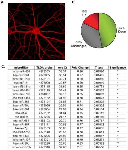
Normal brain development and function depends on microRNA (miRNA) networks to fine tune the balance between the transcriptome and proteome of the cell. These small non-coding RNA regulators are highly enriched in brain where they play key roles in neuronal development, plasticity and disease. In neurodegenerative disorders such as Alzheimer's disease (AD), brain miRNA profiles are altered; thus miRNA dysfunction could be both a cause and a consequence of disease. Our study dissects the complexity of human AD pathology, and addresses the hypothesis that amyloid-beta (Abeta) itself, a known causative factor of AD, causes neuronal miRNA deregulation, which could contribute to the pathomechanisms of AD. We used sensitive TaqMan low density miRNA arrays (TLDA) on murine primary hippocampal cultures to show that about half of all miRNAs tested were down-regulated in response to Abeta peptides. Time-course assays of neuronal Abeta treatments show that Abeta is in fact a powerful regulator of miRNA levels as the response of certain mature miRNAs is extremely rapid. Bioinformatic analysis predicts that the deregulated miRNAs are likely to affect target genes present in prominent neuronal pathways known to be disrupted in AD. Remarkably, we also found that the miRNA deregulation in hippocampal cultures was paralleled in vivo by a deregulation in the hippocampus of Abeta42-depositing APP23 mice, at the onset of Abeta plaque formation. In addition, the miRNA deregulation in hippocampal cultures and APP23 hippocampus overlaps with those obtained in human AD studies. Taken together, our findings suggest that neuronal miRNA deregulation in response to an insult by Abeta may be an important factor contributing to the cascade of events leading to AD.


Conflict of interest statement

Competing Interests: The authors have a purely academic collaboration with Matthias Staufenbiel of Novartis. There is no commercial interest attached to this work with him. All PLoS ONE policies regarding the sharing of data and materials will be adhered to.

Figures

Figure 1
Figure 1. Deregulated miRNAs in mouse primary hippocampal cells treated with Aβ42.
A. Mouse primary hippocampal neurons grown for 24 DIV (days in vitro) stained with neuronal β3 tubulin showing dense axonal networks indicative of healthy mature neurons. Scale bar  = 25 um. B. Neuronal miRNA response to Aβ treatment. Overview of directional miRNA changes after Aβ treatment. miRNAs altered by 15% compared to untreated controls were considered unchanged. C. Summary of significantly deregulated microRNAs in primary hippocampal cells with or without Aβ42 treatment (n = 3) analyzed by rodent TLDA. miRNA expression levels can be gauged using average (Ave) Ct values. T-test P-value significance: **P<0.01, *P<0.05.
Figure 2
Figure 2. miRNA expression pattern in response to Aβ time course in murine primary hippocampal neurons assessed by real-time PCR using TaqMan assays.
Independent primary neuronal preparations treated with Aβ42 for 1, 6 or 15 hours were assayed for miRNA expression relative to untreated controls. T-test P-value significance: ***P<0.001, **P<0.01, *P<0.05. Expression was normalized to snoRNA135.
Figure 3
Figure 3. In vivo analysis of the Aβ plaque-forming APP23 mouse model.
A. In situ hybridization showing APP expression pattern in brains of three month-old APP23 versus wild-type mice. Note the high expression of the transgene in cortex and hippocampus. B. Immunohistochemistry with the 6E10 antibody showing high levels of Aβ in the hippocampus of three month-old APP23 mice compared to littermate controls. Scale bar  = 500 µm. C–E. miRNA expression in hippocampus of two- (C.), seven- (D.) and thirteen- (E.) month-old APP23 mice compared to wild-type littermate controls (n = 4) represented as fold change (FC) as determined using real-time PCR with TaqMan assays. Down-regulated miRNAs have been highlighted in green and up-regulated ones in red. Reactions were normalized to snoRNA135 and P-value significance was calculated using the Students T-test: P-value  = *<0.05, **<0.01, ***<0.001.

Similar articles

Cited by

  • MicroRNA dysregulation in the spinal cord following traumatic injury.
    Yunta M, Nieto-Díaz M, Esteban FJ, Caballero-López M, Navarro-Ruíz R, Reigada D, Pita-Thomas DW, del Águila A, Muñoz-Galdeano T, Maza RM. Yunta M, et al. PLoS One. 2012;7(4):e34534. doi: 10.1371/journal.pone.0034534. Epub 2012 Apr 12. PLoS One. 2012. PMID: 22511948 Free PMC article.
  • MicroRNAs in Neurotoxicity.
    Kaur P, Armugam A, Jeyaseelan K. Kaur P, et al. J Toxicol. 2012;2012:870150. doi: 10.1155/2012/870150. Epub 2012 Mar 6. J Toxicol. 2012. PMID: 22523492 Free PMC article.
  • Bias in microRNA functional enrichment analysis.
    Bleazard T, Lamb JA, Griffiths-Jones S. Bleazard T, et al. Bioinformatics. 2015 May 15;31(10):1592-8. doi: 10.1093/bioinformatics/btv023. Epub 2015 Jan 20. Bioinformatics. 2015. PMID: 25609791 Free PMC article.
  • Differential DNA Methylation of MicroRNA Genes in Temporal Cortex from Alzheimer's Disease Individuals.
    Villela D, Ramalho RF, Silva AR, Brentani H, Suemoto CK, Pasqualucci CA, Grinberg LT, Krepischi AC, Rosenberg C. Villela D, et al. Neural Plast. 2016;2016:2584940. doi: 10.1155/2016/2584940. Epub 2016 Apr 26. Neural Plast. 2016. PMID: 27213057 Free PMC article.
  • A combined miRNA-piRNA signature to detect Alzheimer's disease.
    Jain G, Stuendl A, Rao P, Berulava T, Pena Centeno T, Kaurani L, Burkhardt S, Delalle I, Kornhuber J, Hüll M, Maier W, Peters O, Esselmann H, Schulte C, Deuschle C, Synofzik M, Wiltfang J, Mollenhauer B, Maetzler W, Schneider A, Fischer A. Jain G, et al. Transl Psychiatry. 2019 Oct 7;9(1):250. doi: 10.1038/s41398-019-0579-2. Transl Psychiatry. 2019. PMID: 31591382 Free PMC article.

References

    1. Haass C. Take five–BACE and the gamma-secretase quartet conduct Alzheimer's amyloid beta-peptide generation. EMBO J. 2004;23:483–488. - PMC - PubMed
    1. Theuns J, Brouwers N, Engelborghs S, Sleegers K, Bogaerts V, et al. Promoter mutations that increase amyloid precursor-protein expression are associated with Alzheimer disease. Am J Hum Genet. 2006;78:936–946. - PMC - PubMed
    1. Rovelet-Lecrux A, Hannequin D, Raux G, Le Meur N, Laquerriere A, et al. APP locus duplication causes autosomal dominant early-onset Alzheimer disease with cerebral amyloid angiopathy. Nat Genet. 2006;38:24–26. - PubMed
    1. Podlisny MB, Lee G, Selkoe DJ. Gene dosage of the amyloid beta precursor protein in Alzheimer's disease. Science. 1987;238:669–671. - PubMed
    1. Gotz J, Schild A, Hoerndli F, Pennanen L. Amyloid-induced neurofibrillary tangle formation in Alzheimer's disease: insight from transgenic mouse and tissue-culture models. Int J Dev Neurosci. 2004;22:453–465. - PubMed

Publication types