. 2010 Jun 11;5(6):e11084.
doi: 10.1371/journal.pone.0011084.

Radiotherapy suppresses angiogenesis in mice through TGF-betaRI/ALK5-dependent inhibition of endothelial cell sprouting

Affiliations

Radiotherapy suppresses angiogenesis in mice through TGF-betaRI/ALK5-dependent inhibition of endothelial cell sprouting

Natsuko Imaizumi et al. PLoS One. .

Abstract

Background: Radiotherapy is widely used to treat cancer. While rapidly dividing cancer cells are naturally considered the main target of radiotherapy, emerging evidence indicates that radiotherapy also affects endothelial cell functions, and possibly also their angiogenic capacity. In spite of its clinical relevance, such putative anti-angiogenic effect of radiotherapy has not been thoroughly characterized. We have investigated the effect of ionizing radiation on angiogenesis using in vivo, ex vivo and in vitro experimental models in combination with genetic and pharmacological interventions.

Principal findings: Here we show that high doses ionizing radiation locally suppressed VEGF- and FGF-2-induced Matrigel plug angiogenesis in mice in vivo and prevented endothelial cell sprouting from mouse aortic rings following in vivo or ex vivo irradiation. Quiescent human endothelial cells exposed to ionizing radiation in vitro resisted apoptosis, demonstrated reduced sprouting, migration and proliferation capacities, showed enhanced adhesion to matrix proteins, and underwent premature senescence. Irradiation induced the expression of P53 and P21 proteins in endothelial cells, but p53 or p21 deficiency and P21 silencing did not prevent radiation-induced inhibition of sprouting or proliferation. Radiation induced Smad-2 phosphorylation in skin in vivo and in endothelial cells in vitro. Inhibition of the TGF-beta type I receptor ALK5 rescued deficient endothelial cell sprouting and migration but not proliferation in vitro and restored defective Matrigel plug angiogenesis in irradiated mice in vivo. ALK5 inhibition, however, did not rescue deficient proliferation. Notch signaling, known to hinder angiogenesis, was activated by radiation but its inhibition, alone or in combination with ALK5 inhibition, did not rescue suppressed proliferation.

Conclusions: These results demonstrate that irradiation of quiescent endothelial cells suppresses subsequent angiogenesis and that ALK5 is a critical mediator of this suppression. These results extend our understanding of radiotherapy-induced endothelial dysfunctions, relevant to both therapeutic and unwanted effects of radiotherapy.


Conflict of interest statement

Competing Interests: The authors have declared that no competing interests exist.

Figures

Figure 1
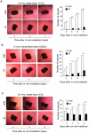
Figure 1. Inhibition of Matrigel plug angiogenesis by skin pre-irradiation.
(a) VEGF-induced Matrigel plug angiogenesis assay was performed within non-irradiated or 20 Gy pre-irradiated areas on the back of distinct C57/BL6 mice. Bar = 0.5 cm. *P<0.05. (n = 7). (b) FGF-2 induced Matrigel plug angiogenesis assay was performed within non-irradiated area either on the back of distinct Swiss nude mice (upper panels) or within and outside a 20 Gy irradiated dorsal area on the same mouse (lower panes) (n = 7). Matrigel without growth factor was used as a negative control. Angiogenesis was quantified by measuring haemoglobin concentration in the recovered Matrigel plugs. Bar = 0.5 cm. *P<0.0001. (c) Staining of CD31 positive endothelial cells (red) and DAPI (blue) was performed on frozen Matrigel sections. Bar = 60 µm. Matrigel implanted within the irradiated area lacks endothelial cells (n = 10). NIR: non-irradiated, IR: irradiated.
Figure 2
Figure 2. Quiescent endothelial cells in vivo and in vitro are resistant to radiation-induced apoptosis.
(a) Sections of non-irradiated skin and of 20 Gy irradiated skin 6 days after irradiation were stained by immunohistochemistry for CD31 (brown). Bar = 60 µm. The graph on the right represents the microvessel density (MVD) of the skin at day 1, 3 and 6 after irradiation and non-irradiated controls. For quantification 5 regions were selected from each slide (n = 5). There was no significant difference in the MVD of irradiated vs. non-irradiated skin. (b) Apoptosis detection in irradiated skin by TUNEL assay. Frozen skin sections prepared before (t = 0), and 4 hours, 24 hours and 10 days after local radiation (20 Gy) were stained with TUNEL reaction (green), anti-CD31 mAb (red) and DAPI (blue). Bars  = x100: 60 µm, x200: 30 µm At 10 days after radiation, keratinocytes within the irradiated skin are largely TUNEL positive, while endothelial cells are negative. The numbers above the images give the quantification of TUNEL positive cells within the total cell population (blue) and CD31+ endothelial cells (red). No TUNEL+ endothelial cells were detected. (c) Detection of apoptosis in irradiated confluent HUVEC cultures. Left panel: frequency of apoptotic cells 4 days after 15 Gy irradiation of confluent HUVEC. Right panel: frequency of apoptotic cells in HUVEC cultures irradiated, split 4 days after radiation and cultured for another 4 days. There was no significant increase in apoptosis within irradiated confluent HUVEC cultures. Apoptotic cells were detected by Annexin V/7AAD staining. NIR: non-irradiated, IR: irradiated.
Figure 3
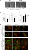
Figure 3. Irradiation induces senescence of confluent endothelial cells.
(a) Confluent HUVEC were irradiated with 15 Gy or not, and 4 days later cultures were split at 1∶3 dilutions and cultures photographed at 1, 3, and 5 days after splitting. Right panel gives the number of cells in non-irradiated and irradiated cultures at the indicated times (n = 5). Bars = 50 µm. (b) Flow cytometry-based cell cycle analysis of confluent HUVEC collected 4 days after radiation. Cells were stained with an anti-Ki67 antibody and 7-AAD dye. Bars show the relative percentage of HUVEC in the different phases of the cell cycle (G0, G1 and G2/M1). (c) Non-irradiated and 15 Gy irradiated HUVEC were fixed 4 or 96 hours after irradiation and stained to detect senescence-associated β-galactosidase activity at pH = 6. Pictures show stained (β-gal staining) and unstained cultures (phase). Irradiated HUVEC acquired β-gal staining and a flattened, enlarged and granular cytoplasm. The bar graph gives the number of β-gal positive cells normalized by total number of cells. (n = 5). *P<0.00001.
Figure 4
Figure 4. Irradiation inhibits endothelial sprouting from aortic rings.
(a) C57/BL6 mice received 15 Gy whole body irradiation. Aorta were explanted at day 5, sliced into rings and embedded in collagen I gels. The number of sprouts was quantified and images were taken at the indicated days after embedding. Aortic rings obtained from non-irradiated mice were used as control. *P<0.01, **P<0.001. (b) C57/BL6 mice received 5 times 3 Gy whole body radiation every 2 days (total dose  = 15 Gy). The aorta was explanted 5 days after the last dose, sliced into rings and embedded in collagen I gels. Aortic rings obtained from non-irradiated mice were used as positive controls. The number of sprouts was quantified and images were taken at the indicated days. Bars = 500 µm. *P<0.0001. NIR: non-irradiated, IR: irradiated. (c) Aortas were explanted from wild type C57/BL6 mice, sliced into rings, embedded in collagen I gels and 2 days later irradiated with 8 Gy. The number of sprouts was quantified immediately before (t = 0), and at 1, 3, and 5 days after radiation. Bars = 500 µm. *P<0.01, **P<0.001. n.s, non significant. For each assay the number of sprouts was counted manually each day under a dissection microscope. Bar graphs on the right represent the number of sprouts. White bars: sprouting from non-irradiated mice or rings; Black bars, sprouting from irradiated mice or rings.
Figure 5
Figure 5. Irradiation inhibits sprouting of isolated endothelial cells.
HUVEC spheroids were embedded in collagen gels and exposed to 8 Gy radiation. The cumulative sprout length was quantified from 10 randomly observed spheroids in each condition 24 hours later. There is a dose-dependent decrease of sprouting in irradiated cultures. Bars = 100 µm. *P<0.01, **P<0.001. NIR: non-irradiated, IR: irradiated.
Figure 6
Figure 6. Irradiation impairs endothelial cell migration.
(a) The effect of radiation on cell migration was examined by a scratch wound closure assay in vitro. Confluent HUVEC were irradiated with 15 Gy, and 4 days later a scratch-wound was created. The micrographs represent the wound immediately after the scratch (t = 0) and at 10 hours later. Bars = 30 µm (b) Wound closure was monitored by time-lapse microscopy and the distance and speed of individual cells were calculated. Ionizing radiations inhibit HUVEC migration. *P<0.0001. (c) Non-irradiated and 15 Gy irradiated HUVEC were tested for adhesion to collagen I and fibronectin on a short-term adhesion assay. *P<0.0001. Coll-I: collagen I, Fn: fibronectin. Irradiated cells adhere more efficiently to both substrates. (d) Radiation effect on adhesion complexes and the cytoskeleton. Confluent HUVEC were irradiated with 15 Gy. Four, 24 hours and 4 days after radiation, a line-scratch wound was made on the cell monolayer, and 10 hours later, cultures were fixed and stained by immunofluorescence with phalloidin (red) and anti-paxillin antibody (green) to reveal the actin cytoskeleton and focal adhesions. Pictures of cells at the wound edge and at confluent region are shown. Migrating NIR cells at the wounding edge are polarized (L, leading edge; T, trailing end), while non-migrating IR cells and cells in confluent regions are not polarized. Bars = 5 µm. NIR: non-irradiated, IR: irradiated conditions.
Figure 7
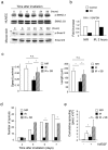
Figure 7. Role of radiation-induced activation of the P53-P21 pathway in senescence and migration.
(a) Confluent HUVEC were irradiated at 15 Gy or not and analyzed for DNA double strand breaks by immunofluorescence staining using anti-P-H2AX mAb (green) at indicated time points. DAPI staining (blue) was used to detect nuclear DNA. Double strand breaks were repaired within 24 hours. (n = 5). Bars = 5 µm. (b) The expression level of three senescence-associated proteins, P16, P21 and P53, was analyzed by Western blotting before (0 hours) and after radiation at indicated time points. Actin was used as loading control. P53 levels are increased 6 hours after radiation, followed by an increase in P21 level. (n = 3). (c) HUVEC were transduced with lentiviruses expressing four different P21shRNA (1–4), no insert (empty vector) and non-silencing shRNA. HUVEC infected with P21shRNA # 3 were used for the functional experiments. (d) Senescence β-gal staining on control and P21 silenced HUVEC at 4 hours and 4 days after irradiation. (n = 6). (e) Migration ability of P21 silenced HUVEC was monitored by wound scratching assay. There was partial rescue of migration defect and decrease in number of senescence positive cells in P21 silenced HUVEC. (n = 3) *P<0.001. NIR: non-irradiated, IR: irradiated, NS: non-silencing HUVEC.
Figure 8
Figure 8. Radiation activates the TGF-βRI/ALK5 pathway and ALK5 inhibition prevents radiation-induced suppression of migration and sprouting.
(a) Increased Smad2 phosphorylation in irradiated cultured HUVEC and mouse skin (15 Gy and 20 Gy respectively) demonstrated by Western blotting analysis. Induction of p-Smad2 was biphasic with early peaks at 2–6 hours and late peaks at 24–96 hours. (n = 3). (b) Radiation induces the TGF-β pathway target PAI-1 gene in HUVEC. HUVEC were treated with the ALK5 inhibitor SB431542 at 10 µM one day before radiation. RNA was extracted before and 2 hours after irradiation and PAI-1 mRNA quantified by real time RT-PCR. (c) The ALK5 inhibitor SB431542 (SB) rescued the migration defects caused by radiation. Left panel: migration speed; right panel: migration distance. (n = 10) *P<0.001. (d) The ALK5 inhibitor SB431542 (SB) rescued the sprouting defects caused by radiation. The SB compound was added in the medium during the whole mouse aortic ring assay. Level of endothelial cell sprouting in the presence of SB reached to levels observed in non-irradiated/non-treated rings (n = 9) *P<0.05. (e) The ALK5 inhibitor SB431542 (SB) rescued the radiation-induced sprouting defects in HUVEC spheroid assay. SB compound was added to spheroid-containing collagen gel at 10 µM and the spheroids were exposed to 15 Gy radiation. Endothelial sprouting was quantified 24 hours after incubation. (n = 10) *P<0.05, **P<0.001. NIR: non-irradiated, IR: irradiated, SB: SB431542.
Figure 9
Figure 9. The ALK5 inhibitor SB431542 prevents radiation-induced inhibition of angiogenesis in the Matrigel plug assay.
VEGF-induced angiogenesis was performed in non-irradiated or irradiated (20 Gy) C57/BL6 mice, which were treated with SB431542 (SB) compound, or vehicle only, daily from one day before irradiation until the end of the experiment. One week after Matrigel implantation, angiogenesis was quantified by measuring haemoglobin content. Representative images of recovered plugs and quantification of haemoglobin content are shown. (n = 7). Bars = 0.5 cm. *P<0.005. n.s., non significant. NIR: non-irradiated, IR: irradiated: SB: SB431542.

References

    1. Bernier J, Hall EJ, Giaccia A. Radiation oncology: a century of achievements. Nat Rev Cancer. 2004;4:737–747. - PubMed
    1. Rosen EM, Fan S, Rockwell S, Goldberg ID. The molecular and cellular basis of radiosensitivity: implications for understanding how normal tissues and tumors respond to therapeutic radiation. Cancer Invest. 1999;17:56–72. - PubMed
    1. Barcellos-Hoff MH, Park C, Wright EG. Radiation and the microenvironment - tumorigenesis and therapy. Nat Rev Cancer. 2005;5:867–875. - PubMed
    1. Garcia-Barros M, Paris F, Cordon-Cardo C, Lyden D, Rafii S, et al. Tumor response to radiotherapy regulated by endothelial cell apoptosis. Science. 2003;300:1155–1159. - PubMed
    1. Fajardo LF. The pathology of ionizing radiation as defined by morphologic patterns. Acta Oncol. 2005;44:13–22. - PubMed

Publication types

MeSH terms

Substances