. 2009:41:167-218.
doi: 10.1016/S0065-2601(08)00404-8.

Affect as a Psychological Primitive

Affiliations

Affect as a Psychological Primitive

Lisa Feldman Barrett et al. Adv Exp Soc Psychol. 2009.

Abstract

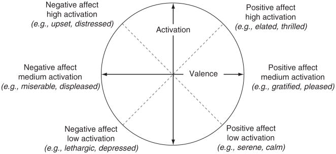
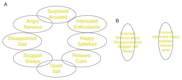
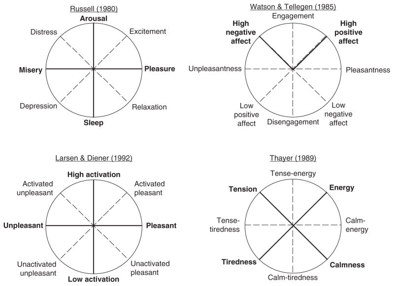
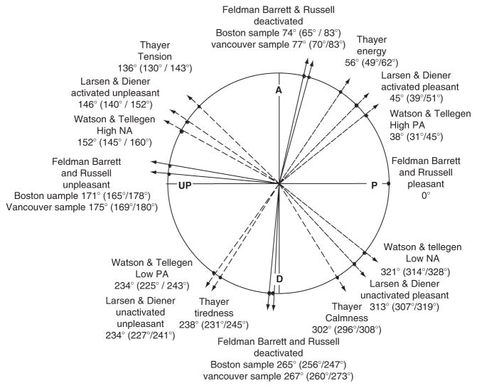
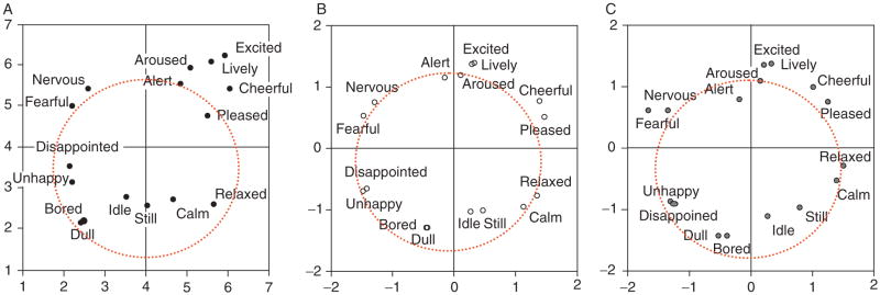
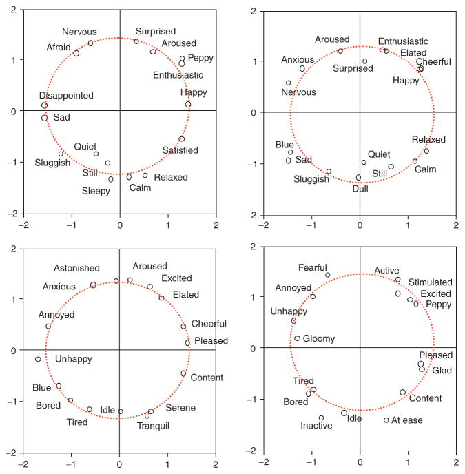
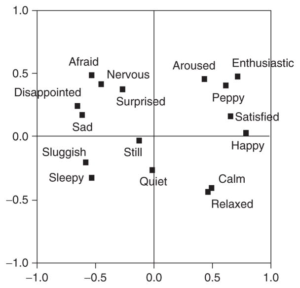
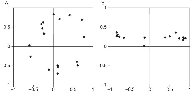
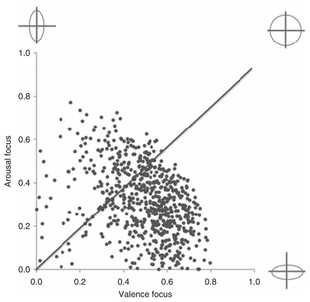
In this article, we discuss the hypothesis that affect is a fundamental, psychologically irreducible property of the human mind. We begin by presenting historical perspectives on the nature of affect. Next, we proceed with a more contemporary discussion of core affect as a basic property of the mind that is realized within a broadly distributed neuronal workspace. We then present the affective circumplex, a mathematical formalization for representing core affective states, and show that this model can be used to represent individual differences in core affective feelings that are linked to meaningful variation in emotional experience. Finally, we conclude by suggesting that core affect has psychological consequences that reach beyond the boundaries of emotion, to influence learning and consciousness.


Figures

Figure 4.1
Figure 4.1
The hypothesized neural reference space for core affect. Brain areas that realize core affect include the visceromotor and sensory integration networks in the OFC (A–C, blue, and purple, respectively), the anterior insula (D, yellow), the amygdala (D, rose), subgenual and pregenual parts of the ACC (B, copper, tan), the hypothalamus (B, light green), and the ventral striatum (D, dark green). Also included are the midbrain (B, turquoise) and brainstem (B, C, dark pink). Adapted from Barrett et al. (2007). Refer online version of the chapter for color figure.
Figure 4.2
Figure 4.2
The observed neural reference space for core affect. 165 neuroimaging studies of emotion (58 using PET and 107 using fMRI) published from 1990 to 2005 were summarized in a multilevel meta-analysis to produce the observed neural reference space for emotion (Wager et al., 2008). These areas include (from top left, clockwise) anterior insula (aIns), lateral OFC (lOFC), pregenual cingulate cortex (pgACC), subgenual cingulate cortex (sgACC), ventral medial prefrontal cortex (vmPFC), temporal cortex/amygdala (TC/Amygdala), thalamus, ventral striatrum (v Striatum), nucleus accumbens, hypothalamus, midbrain, pons, medulla, OFC, and basal forebrain. Other areas shown in this figure (e.g., inferior frontal gyrus (IFG), superior temporal cortex (sTC), dorsal medial prefrontal cortex (dmPFC), posterior cingulate cortex (PCC), medial temporal cortex (mTC), and cerebellum (CB)) relate to other psychological processes involved with emotion perception and experience. (See online version of the chapter for color figure).
Figure 4.3
Figure 4.3
The affective circumplex. Hedonic valence is represented on the horizontal axis and arousal on the vertical axis.
Figure 4.4
Figure 4.4
Variations in the affective circumplex. (A) depicts a prototypical affective space insofar as emotions are distributed evenly in a circular structure, with many smaller regions of homogeneity, where each region is psychologically distinct from every other. (B) depicts a nonprototypical affective space with two larger regions, where emotions within a region are highly similar. Figure is adapted from Barrett (2004).
Figure 4.5
Figure 4.5
Multiple affective dimensions mapped in circumplex space. Primary (or main) dimensions are indicated in with black solid lines and labeled with capital letters. Secondary dimensions are indicated with gray dotted lines and are labeled in lower case letters. From Barrett and Russell (1999).
Figure 4.6
Figure 4.6
A circumplex representation of various affective dimensions plotted according to a CIRCUM analysis. The Russell/Barrett, Larsen/Diener, Thayer, and Watson/Tellegen affective dimensions were measured using separate scales and there position in circular space was estimated using a structural equation modeling program (CIRCUM) that was specifically designed to estimate circumplexity. From Yik et al. (1999).
Figure 4.7
Figure 4.7
The “Necker” cube illusion.
Figure 4.8
Figure 4.8
Brain areas consistently activated for positive (yellow) and negative (blue) affective experiences. OFC = orbitofrontal cortex; vaINS = ventral anterior insula; Amy = amygdala; vStr = ventral striatum; vGP = ventral globus pallidus; pgACC = pregenual anterior cingulated cortex; rdACC = rostral dorsal anterior cingulate cortex; vmPFC = ventromedial prefrontal cortex; Hy = hypothalamus; Thal = thalamus; PAG/SC = periaquaductal gray/superior colliculus; aINS = anterior insular. From Wager et al. (2008). (See online version of the chapter for color figure).
Figure 4.9
Figure 4.9
Cognitive maps of affective space. The circumplex structure of affect derived from direct semantic ratings, similarity judgments, and conditional probability judgments of emotion words. Based on data from Barrett and Fossum (2001).
Figure 4.10
Figure 4.10
Affective circumplex structures estimated from multidimensional scalings of similarity judgments using different sets of emotion adjectives (Barrett, unpublished data).
Figure 4.11
Figure 4.11
Cross-sectional ratings of emotional experience modeled as a circumplex. Factor loading plot for ratings of emotional experience taken using 16 adjectives. Valence is represented as the horizontal axis and arousal as the vertical axis. Taken from Feldman (1995b).
Figure 4.12
Figure 4.12
Idiographic variation in circumplex structure. Examples of idiographic affective circumplexes derived from momentary ratings of emotional experience for two participants. The participant depicted in (A) has a relatively prototypical circumplex with many small regions of homogeneity which reflects high emotional granularity. The participant in (B) has a flatter, more elliptically shaped circumplex which reflects low emotional granularity. Figure reprinted from Feldman (1995b).
Figure 4.13
Figure 4.13
Scatterplot of variation in valence focus and arousal focus. Valence focus plotted against arousal focus for ~700 subjects who completed experience sampling experiments in our laboratory over a 10-year period. Caricatured circumplex structures are plotted on the extremes of the axes. Participants who fall along the diagonal line (where VF = AF) are high in emotional granularity and have a prototypical circumplex structure. Participants who fall above the diagonal like (AF > VF) and below the diagonal line (VF > AF) are less granular and have more elliptical shaped circumplex structures.
Figure 4.14
Figure 4.14
Variation in affective learning. Sympathetic nervous system response (as indexed by EDA) to face stimuli that were either consistently or were never paired with an aversive electric shock during an associative affective learning paradigm (A). The stimulus that was paired with a shock (CS+) acquired affective value as indicated by a significantly higher EDA response as compared with the EDA response to the stimulus that was never paired with shock (CS−). Individual differences in the acquisition of affective value were related to variation in affective reactivity (B). The relationship between perceptual sensitivity to affective value and the magnitude of affective learning is presented at three levels of neuroticism. From Bliss-Moreau et al. (manuscript under review).
Figure 4.15
Figure 4.15
Individual differences in rule-based affective learning. Positive affective learning via rule-based means is predicted by participants’ sensitivity to positive information and propensity to experience positive affect (as indexed by self-reported extra-version). Adapted from Bliss-Moreau et al. (2008, Study 3).

Similar articles

Cited by

References

    1. Abelson RP, Sermat V. Multidimensional scaling of facial expressions. Journal of Experimental Psychology. 1962;63:546–554. - PubMed
    1. Alpers GW, Gerdes ABM. Here is looking at you: Emotional faces predominate in binocular rivalry. Emotion. 2007;7:495–506. - PubMed
    1. Alpers GW, Pauli P. Emotional pictures predominate in binocular rivalry. Cognition and Emotion. 2006;20:596–607.
    1. Alpers GW, Ruhleder M, Walz N, Mühlberger A, Pauli P. Binocular rivalry between emotional and neutral stimuli: A validation using fear conditioning and EEG. International Journal of Psychophysiology. 2005;57:25–32. - PubMed
    1. Arnold M. Emotion and personality. New York: Columbia University Press; 1960.

LinkOut - more resources