Review
. 2010 Aug;117(8):981-1000.
doi: 10.1007/s00702-010-0429-0. Epub 2010 Jun 15.

Motor neuron-immune interactions: the vicious circle of ALS

Affiliations
Review

Motor neuron-immune interactions: the vicious circle of ALS

Ana G Barbeito et al. J Neural Transm (Vienna). 2010 Aug.

Abstract

Because microglial cells, the resident macrophages of the CNS, react to any lesion of the nervous system, they have for long been regarded as potential players in the pathogenesis of several neurodegenerative disorders including amyotrophic lateral sclerosis, the most common motor neuron disease in the adult. In recent years, this microglial reaction to motor neuron injury, in particular, and the innate immune response, in general, has been implicated in the progression of the disease, in mouse models of ALS. The mechanisms by which microglial cells influence motor neuron death in ALS are still largely unknown. Microglial activation increases over the course of the disease and is associated with an alteration in the production of toxic factors and also neurotrophic factors. Adding to the microglial/macrophage response to motor neuron degeneration, the adaptive immune system can likewise influence the disease process. Exploring these motor neuron-immune interactions could lead to a better understanding in the physiopathology of ALS to find new pathways to slow down motor neuron degeneration.


Figures
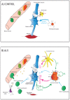
Fig. 1
Fig. 1
Immune cells of the CNS including microglia and T lymphocytes affect motor neuron survival. a Control situation: the presynaptic neuron releases glutamate (Glu), which binds to glutamate receptors (GluRs) on the postsynaptic motor neuron resulting in excitation through calcium influx. Extracellular glutamate is quickly cleared from the synaptic cleft by astrocytes through EAAT2 transporters. In a non-inflamed environment, microglial cells remain in a resting or “surveying” state, most likely releasing factors with neurotrophic influence (blue arrows) b ALS situation: decreased expression of astrocytic glutamate transporters EAAT2 could lead to prolonged glutamate excitation of motor neurons and participate in their degeneration (excitotoxic hypothesis). Activated microglial cells and astrocytes produce toxic factors (pink arrows). Among the factors released by astrocytes, macrophage- colony stimulating Factor (M-CSF) and monocyte chemoattractant protein-1 (MCP-1) are capable of activating microglial cells, increasing their proliferation (M-CSF) or migration (MCP-1) (green arrows). Microglial cells are also prone to self-activation by releasing Tumor Necrosis Factor-α (TNFα) for which they express the receptors 1 and 2 (TNFR1/2) and M-CSF acting on the receptor fms. Activated microglial cells will produce more reactive oxygen species (ROS) like nitric oxide (NO) through the inducible NO synthase and superoxide (O2·−) through activation of NADPH oxidases (Nox1/2), but also proinflammatory cytokines like interleukines 1β and 6 (IL-1β, IL-6) and prostaglandins (PGE2) through activation of the cyclooxygenase 2 (COX2). Extracellular ATP, likely coming from damaged motor neurons, binds to microglial purinergic P2 receptors, therefore, contributing to microglial activation. Motor neurons can also participate to glial cell activation by releasing mutant SOD1 co-secreted with chromogranine (Cg) that can bind to CD14 acting in concert with the Toll-like receptors (TLR2/4). The adaptive immune system is also part of the degenerating motor neuron response. T lymphocytes (CD4+ and CD8+) coming from the periphery enter the spinal cord during the inflammatory process in ALS. CD4+ lymphocytes seem to have a neuroprotective effect by directly releasing anti-inflammatory factors like interleukines 4 and 10 (IL-4, IL-10) or by acting on microglial cells to increase their neurotrophic function (production of insulin-like growth factor-1 (IGF-1)). The role of infiltrating CD8+ T cells remains unclear and B cells are not present in ALS spinal cords. Dendritic (antigen-presenting) cells secrete MCP-1, which probably participates in the infiltration of peripheric immune cells. All together, the inflammatory environment and increased oxidative stress take part in the degeneration of the motor neurons that leads to muscle atrophy in ALS.

References

    1. Ajami B, Bennett JL, Krieger C, Tetzlaff W, Rossi FM. Local self-renewal can sustain CNS microglia maintenance and function throughout adult life. Nat Neurosci. 2007;10:1538–1543. - PubMed
    1. Alexianu ME, Kozovska M, Appel SH. Immune reactivity in a mouse model of familial ALS correlates with disease progression. Neurology. 2001;57:1282–1289. - PubMed
    1. Almer G, Vukosavic S, Romero N, Przedborski S. Inducible nitric oxide synthase up-regulation in a transgenic mouse model of familial amyotrophic lateral sclerosis. J Neurochem. 1999;72:2415–2425. - PubMed
    1. Almer G, Guegan C, Teismann P, Naini A, Rosoklija G, Hays AP, Chen C, Przedborski S. Increased expression of the pro-inflammatory enzyme cyclooxygenase-2 in amyotrophic lateral sclerosis. Ann Neurol. 2001;49:176–185. - PubMed
    1. Andersson PB, Perry VH, Gordon S. The kinetics and morphological characteristics of the macrophage-microglial response to kainic acid-induced neuronal degeneration. Neuroscience. 1991;42:201–214. - PubMed

Publication types

MeSH terms