

Three-dimensional elastic image registration based on strain energy minimization: application to prostate magnetic resonance imaging
- PMID: 20552248
- PMCID: PMC3138929
- DOI: 10.1007/s10278-010-9306-5
Three-dimensional elastic image registration based on strain energy minimization: application to prostate magnetic resonance imaging
Abstract
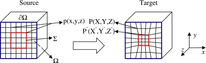
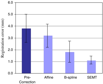
The use of magnetic resonance (MR) imaging in conjunction with an endorectal coil is currently the clinical standard for the diagnosis of prostate cancer because of the increased sensitivity and specificity of this approach. However, imaging in this manner provides images and spectra of the prostate in the deformed state because of the insertion of the endorectal coil. Such deformation may lead to uncertainties in the localization of prostate cancer during therapy. We propose a novel 3-D elastic registration procedure that is based on the minimization of a physically motivated strain energy function that requires the identification of similar features (points, curves, or surfaces) in the source and target images. The Gauss-Seidel method was used in the numerical implementation of the registration algorithm. The registration procedure was validated on synthetic digital images, MR images from prostate phantom, and MR images obtained on patients. The registration error, assessed by averaging the displacement of a fiducial landmark in the target to its corresponding point in the registered image, was 0.2 ± 0.1 pixels on synthetic images. On the prostate phantom and patient data, the registration errors were 1.0 ± 0.6 pixels (0.6 ± 0.4 mm) and 1.8 ± 0.7 pixels (1.1 ± 0.4 mm), respectively. Registration also improved image similarity (normalized cross-correlation) from 0.72 ± 0.10 to 0.96 ± 0.03 on patient data. Registration results on digital images, phantom, and prostate data in vivo demonstrate that the registration procedure can be used to significantly improve both the accuracy of localized therapies such as brachytherapy or external beam therapy and can be valuable in the longitudinal follow-up of patients after therapy.
Figures


Similar articles
-
Mapping of the prostate in endorectal coil-based MRI/MRSI and CT: a deformable registration and validation study.Med Phys. 2004 Nov;31(11):3087-94. doi: 10.1118/1.1806292. Med Phys. 2004. PMID: 15587662 Clinical Trial.
-
Elastic registration of prostate MR images based on estimation of deformation states.Med Image Anal. 2015 Apr;21(1):87-103. doi: 10.1016/j.media.2014.12.007. Epub 2015 Jan 8. Med Image Anal. 2015. PMID: 25624044
-
Use and uncertainties of mutual information for computed tomography/ magnetic resonance (CT/MR) registration post permanent implant of the prostate.Med Phys. 2005 Feb;32(2):473-82. doi: 10.1118/1.1851920. Med Phys. 2005. PMID: 15789594 Clinical Trial.
-
Challenges in accurate registration of 3-D medical imaging and histopathology in primary prostate cancer.Eur J Nucl Med Mol Imaging. 2013 Jul;40 Suppl 1(0 1):S72-8. doi: 10.1007/s00259-013-2382-2. Epub 2013 Mar 16. Eur J Nucl Med Mol Imaging. 2013. PMID: 23503575 Free PMC article. Review.
-
Interactive Feature Space Explorer© for multi-modal magnetic resonance imaging.Magn Reson Imaging. 2015 Jul;33(6):804-15. doi: 10.1016/j.mri.2015.03.007. Epub 2015 Apr 11. Magn Reson Imaging. 2015. PMID: 25868623 Free PMC article. Review.
Cited by
-
Automated, foot-bone registration using subdivision-embedded atlases for spatial mapping of bone mineral density.J Digit Imaging. 2013 Jun;26(3):554-62. doi: 10.1007/s10278-012-9524-0. J Digit Imaging. 2013. PMID: 23090209 Free PMC article.
-
Image guidance for focal therapy of prostate cancer.World J Urol. 2010 Dec;28(6):727-34. doi: 10.1007/s00345-010-0604-9. Epub 2010 Oct 21. World J Urol. 2010. PMID: 20963422 Review.
-
Multi-Task Learning for Registering Images With Large Deformation.IEEE J Biomed Health Inform. 2021 May;25(5):1624-1633. doi: 10.1109/JBHI.2020.3016699. Epub 2021 May 11. IEEE J Biomed Health Inform. 2021. PMID: 32795972 Free PMC article.
References
-
- Sanchez-Chapado M, Angulo JC, Ibarburen C, Aguado F, Ruiz A, Viano J, Garcia-Segura JM, Gonzalez-Esteban J, Rodriquez-Vallejo JM. Comparison of digital rectal examination, transrectal ultrasonography, and multicoil magnetic resonance imaging for preoperative evaluation of prostate cancer. Eur Urol. 1997;21:140–149. - PubMed
-
- Nguyen PL, Chen MH, D’Amico AV, Tempany CM, Steele GS, Albert M, Cormack RA, Carr-Locke DL, Bleday R, Suh WW. Magnetic resonance image-guided salvage brachytherapy after radiation in select men who initially presented with favorable-risk prostate cancer: a prospective phase 2 study. Cancer. 2007;110(7):1485–1492. doi: 10.1002/cncr.22934. - DOI - PubMed
-
- Schnall MD, Imai Y, Tomaszewski J, Pollack HM, Lenkinski RE, Kressel HY. Prostate cancer: local staging with endorectal surface coil MR imaging. Radiology. 1991;178:797–802. - PubMed
Publication types
MeSH terms
LinkOut - more resources
Full Text Sources
Other Literature Sources
Medical
