

Mechanisms of failed apoptotic cell clearance by phagocyte subsets in cardiovascular disease
- PMID: 20552278
- PMCID: PMC3744319
- DOI: 10.1007/s10495-010-0516-6
Mechanisms of failed apoptotic cell clearance by phagocyte subsets in cardiovascular disease
Abstract
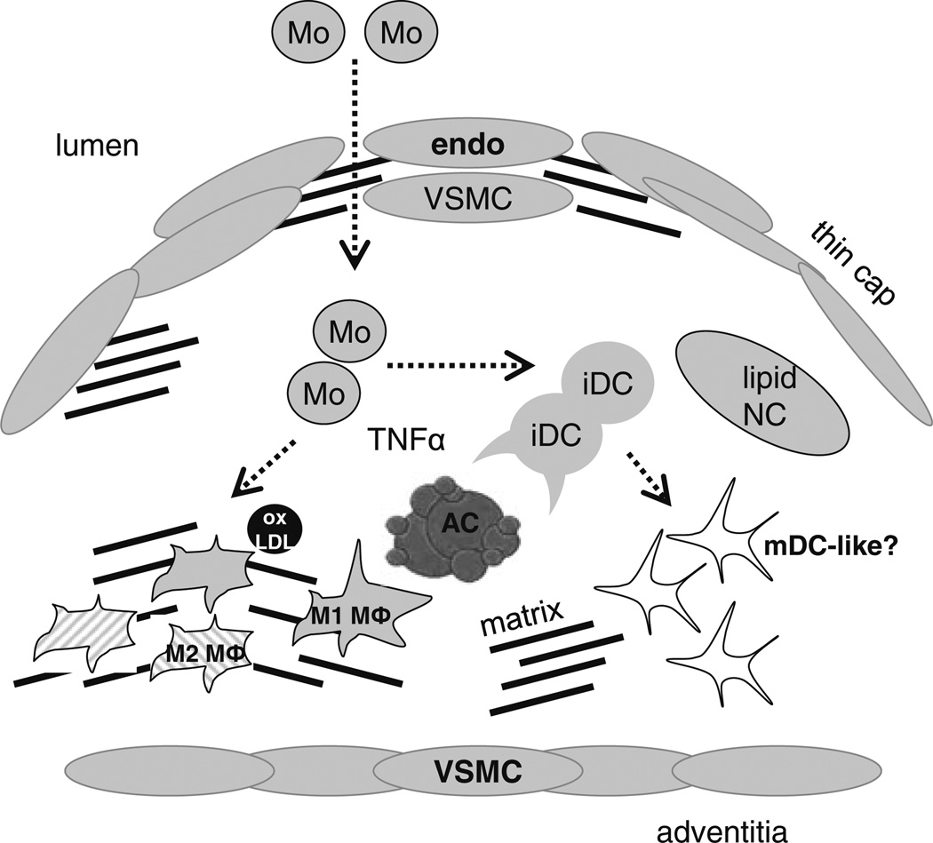
Recent evidence in humans indicate that defective phagocytic clearance of dying cells is linked to progression of advanced atherosclerotic lesions, the precursor to atherothrombosis, ischemic heart disease, and leading cause of death in the industrialized world. During atherogenesis, apoptotic cell turnover in the vascular wall is counterbalanced by neighboring phagocytes with high clearance efficiency, thereby limiting cellularity and maintaining lesion integrity. However, as lesions mature, phagocytic removal of apoptotic cells (efferocytosis) becomes defective, leading to secondary necrosis, expansion of plaque necrotic cores, and susceptibility to rupture. Recent genetic causation studies in experimental rodents have implicated key molecular regulators of efferocytosis in atherosclerotic progression. These include MER tyrosine kinase (MERTK), milk fat globule-EGF factor 8 (MFGE8), and complement C1q. At the cellular level, atheromata are infiltrated by a heterogenous population of professional phagocytes, comprised of monocytes, differentiated macrophages, and CD11c(+) dendritic-like cells. Each cell type is characterized by disparate clearance efficiencies and varying activities of key phagocytic signaling molecules. It is in this context that we outline a working model whereby plaque necrosis and destabilization is jointly promoted by (1) direct inhibition of core phagocytic signaling pathways and (2) expansion of phagocyte subsets with poor clearance capacity. Towards identifying targets for promoting efficient apoptotic cell clearance and resolving inflammation in atherosclerosis and during ischemic heart disease and post myocardial infarction, this review will discuss potential in vivo suppressors of efferocytosis at each stage of clearance and how these putative interventional targets may differentially affect uptake at the level of vascular phagocyte subsets.
Figures


References
-
- Libby P, Nahrendorf M, Pittet MJ, Swirski FK. Diversity of denizens of the atherosclerotic plaque: not all monocytes are created equal. Circulation. 2008;117:3168–3170. - PubMed
-
- Tabas I. Apoptosis and plaque destabilization in atherosclerosis: the role of macrophage apoptosis induced by cholesterol. Cell Death Differ. 2004;11(1):S12–S16. - PubMed
-
- Gregory C. Cell biology: sent by the scent of death. Nature. 2009;461:181–182. - PubMed
-
- Henson PM. Dampening inflammation. Nat Immunol. 2005;6:1179–1181. - PubMed
Publication types
MeSH terms
Grants and funding
LinkOut - more resources
Full Text Sources
Other Literature Sources
Research Materials
Miscellaneous
