. 2010 Oct;468(10):2695-703.
doi: 10.1007/s11999-010-1421-6. Epub 2010 Jun 15.

In vivo and in vitro analysis of rat lumbar spine mechanics

Affiliations

In vivo and in vitro analysis of rat lumbar spine mechanics

Matthew E Cunningham et al. Clin Orthop Relat Res. 2010 Oct.

Abstract

Background: Rodent lumbar and caudal (tail) spine segments provide useful in vivo and in vitro models for human disc research. In vivo caudal models allow characterization of the effect of static and dynamic loads on disc mechanics of individual animals with time, but the lumbar models have required sacrifice of the animals for in vitro mechanical testing.

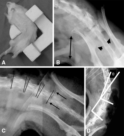
Questions/purposes: We therefore developed a novel displacement controlled in vivo lumbar spine noninvasive induced angular displacement (NIAD) test; data obtained with NIAD were used to compare angular displacement between segmental levels (L4/L5, L5/L6 and L6/S1), interobserver radiograph measurement agreement, and intraobserver radiograph measurement repeatability. Measurements from NIAD were compared with angular displacement, bending stiffness, and moment to failure measured by an in vitro test.

Methods: Anesthetized Lewis rats were xrayed in a 90° angled fixture, and NIAD was measured at lumbar levels L4 to S1 by two independent and blinded observers. After euthanasia, in vitro angular displacement (IVAD), stiffness, and failure moment were measured for the combined L4-L6 segment in four-point bending.

Results: NIAD was greater at L4/L5 and L5/L6 than at L6/S1. Combined coronal NIAD for L4-L6 was 42.8° ± 5.3° and for IVAD was 61.5° ± 3.8°. Reliability assessed by intraclass correlation coefficient (ICC) was 0.905 and 0.937 for intraobserver radiograph measurements, and interobserver ICCs ranged from 0.387 to 0.653 for individual levels. The interobserver ICC was 0.911 for combined data from all levels. Reliability for test-retest NIAD measurements had an ICC of 0.932. In vitro failure moment correlated with NIAD left bending.

Conclusions: The NIAD method yielded reproducible and reliable rat lumbar spine angular displacement measurements without required euthanasia, and allows repetitive monitoring of animals with time. For lumbar spine research studies performed during a course of time, the NIAD method may reduce animal numbers required by providing serial angular displacement measurements without euthanasia.

Clinical relevance: Improved methods to assess comparative models for disease or aging may permit enhanced clinical treatments and improved patient care.


Figures

Fig. 1
Fig. 1
A flowchart for the study design is depicted. The left column shows how interobserver and intraobserver radiographic measurements were assessed, and where the ANOVA assessments were made to compare segmental angular displacement for each observer and between observers. The right column shows how specimens were used for the test-retest, paired in vivo/in vitro, and in vitro biomechanics assessments.
Fig. 2A–D
Fig. 2A–D
(A) An anesthetized Lewis rat is shown in the bending bracket positioned for right-bending radiograph. (B) This high-definition digital radiograph shows inappropriate positioning of the animal in the bracket for right bend assessment with lack of correct force application on the iliac wing (double-ended arrow) and unacceptable rotation judged by the asymmetry of the posterior inferior iliac spines (arrowheads). (C) Bony landmarks for the pelvic measurements (arrows point to the bilateral sacral ala sulci, the most caudal location of the proximal sacral ridge) and vertebral end plate line placement (lines) are shown. The animal is well positioned with symmetric posterior inferior iliac spines and good contact of the iliac wing with the positioning bracket. (D) Cobb end plate measurement (θ) for the L4/L5 segment is shown.
Fig. 3A−D
Fig. 3A−D
(A) This photograph of the four-point bending fixture shows the specimen in maximal deflection in left lateral bending with angular deformation (θ) indicated. Displacement is in the downgoing vertical direction and the load cell is beneath the bottom fixture. (B) Continuous cycling data for a sample tested in flexion are shown. Displacement is graphed as decreasing from right to left during cycling reflecting the apparatus construction and applied force is negative by convention. (C) Load-displacement traces for a representative sample in the fifth loading cycle for each of the specified directions are shown. Stiffness is the slope of curve between 2 and 4 N in the loading phase. The plots for each of the directions were superposed and are illustrated after translation to avoid overlap. (D) A representative bending to failure curve is shown; the arrow marks the failure load (20 N).

References

    1. Alini M, Eisenstein SM, Ito K, Little C, Kettler AA, Masuda K, Melrose J, Ralphs J, Stokes I, Wilke HJ. Are animal models useful for studying human disc disorders/degeneration? Eur Spine J. 2008;17:2–19. doi: 10.1007/s00586-007-0414-y. - DOI - PMC - PubMed
    1. Beckstein JC, Sen S, Schaer TP, Vresilovic EJ, Elliott DM. Comparison of animal discs used in disc research to human lumbar disc: axial compression mechanics and glycosaminoglycan content. Spine (Phila Pa 1976) 2008;33:E166–173. - PubMed
    1. Bettini N, Girardo M, Dema E, Cervellati S. Evaluation of conservative treatment of non specific spondylodiscitis. Eur Spine J. 2009;18(suppl 1):143–150. doi: 10.1007/s00586-009-0979-8. - DOI - PMC - PubMed
    1. Boxberger JI, Auerbach JD, Sen S, Elliott DM. An in vivo model of reduced nucleus pulposus glycosaminoglycan content in the rat lumbar intervertebral disc. Spine (Phila Pa 1976) 2008;33:146–154. - PMC - PubMed
    1. Boxberger JI, Orlansky AS, Sen S, Elliott DM. Reduced nucleus pulposus glycosaminoglycan content alters intervertebral disc dynamic viscoelastic mechanics. J Biomech. 2009;42:1941–1946. doi: 10.1016/j.jbiomech.2009.05.008. - DOI - PMC - PubMed