Review
. 2010 Oct;17(5):897-912.
doi: 10.1007/s12350-010-9263-x.

Molecular imaging of inflammation and intraplaque vasa vasorum: a step forward to identification of vulnerable plaques?

Affiliations
Review

Molecular imaging of inflammation and intraplaque vasa vasorum: a step forward to identification of vulnerable plaques?

Gerrit L ten Kate et al. J Nucl Cardiol. 2010 Oct.

Abstract

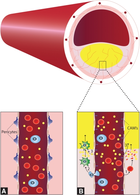
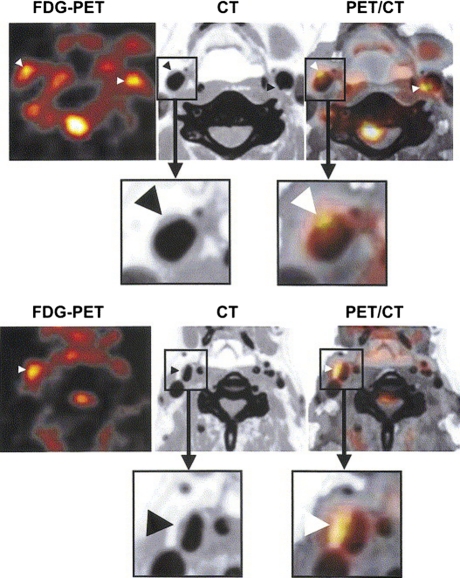
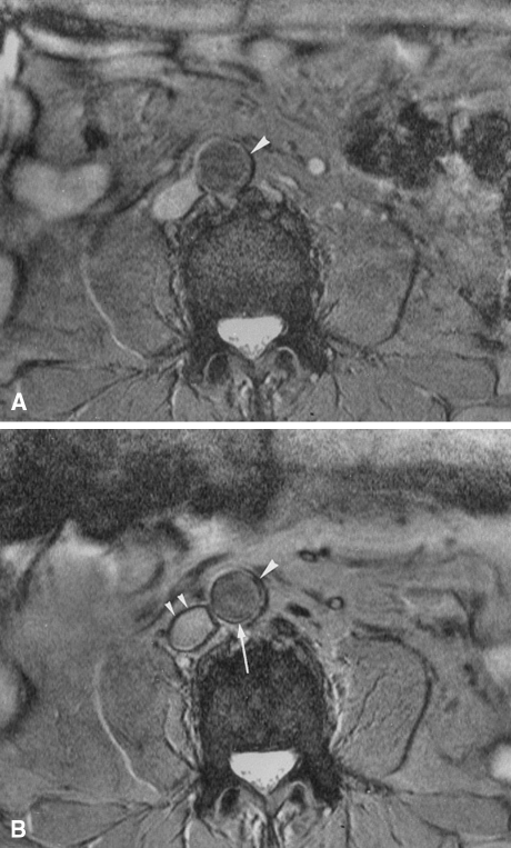
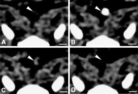
Current developments in cardiovascular biology and imaging enable the noninvasive molecular evaluation of atherosclerotic vascular disease. Intraplaque neovascularization sprouting from the adventitial vasa vasorum has been identified as an independent predictor of intraplaque hemorrhage and plaque rupture. These intraplaque vasa vasorum result from angiogenesis, most likely under influence of hypoxic and inflammatory stimuli. Several molecular imaging techniques are currently available. Most experience has been obtained with molecular imaging using positron emission tomography and single photon emission computed tomography. Recently, the development of targeted contrast agents has allowed molecular imaging with magnetic resonance imaging, ultrasound and computed tomography. The present review discusses the use of these molecular imaging techniques to identify inflammation and intraplaque vasa vasorum to identify vulnerable atherosclerotic plaques at risk of rupture and thrombosis. The available literature on molecular imaging techniques and molecular targets associated with inflammation and angiogenesis is discussed, and the clinical applications of molecular cardiovascular imaging and the use of molecular techniques for local drug delivery are addressed.


Figures

Figure 1
Figure 1
Schematic cross section of an atherosclerotic vessel with a large plaque, lipid core, and intraplaque vasa vasorum sprouting from the adventitial vasa vasorum network. Panel A shows the normal structure of the vasa vasorum with tight gap junctions. Panel B shows newly formed vasa vasorum with increased endothelial gap junctions, pericyte loss and expression of cellular adhesion molecules, the extravasation of erythrocytes and their consequent destruction, the adhesion and extravasation of monocytes and their subsequent transformation to macrophages and resultant foam cells and the direct extravasation of lipids. These factors cause growth of the lipid core of the atherosclerotic plaque
Figure 2
Figure 2
Transaxial images of FDG-PET (left), CT (middle), and co-registration of PET and CT (right) showing FDG uptakes in the carotid arterial plaques (arrowheads) of two patients. Reproduced with permission from Elsevier
Figure 3
Figure 3
Axial 2D gradient-echo MRI of a patient. Pre- (A) and postcontrast (B) images display the level of the aorta. On the precontrast image, the aortic wall is homogeneously hyperintense (A, arrowhead). Following SPIO administration, a pronounced signal loss of an area extending from the inner to the outer surface of the aortic wall can be seen (B, arrowhead). This vessel segment was considered positive. Note, however, that there is also a low-intensity ring at the interface between the aortic wall and lumen on the postcontrast image (B, long arrow). It is not possible to precisely determine whether the ring is truly confined to the aortic wall or to the lumen. This appearance is defined as a ring phenomenon, which is also seen in other vessels, such as the inferior vena cava (B, small arrowheads). Reproduced with permission from John Wiley & Sons
Figure 4
Figure 4
Axial views of the same atherosclerotic plaque (white arrowheads) in the aorta of a rabbit, obtained by CT before (A), during (B) and 2 h after the injection of an iodine nanoparticle contrast agent (C) or a conventional iodine contrast agent (D). Adapted with permission from Macmillan Publishers Ltd
Figure 5
Figure 5
Transcutaneous ultrasound images of the atherosclerotic left carotid artery of a Yucatan miniswine. After injection of saline (A). After injection of nontargeted microbubbles (B) (arrows point to microbubbles within the lumen). After injection of ICAM-1 targeted microbubbles (C) (arrows point to microbubbles attached to the atherosclerotic plaque). Reproduced with permission from Elsevier

References

    1. Weissleder R, Pittet MJ. Imaging in the era of molecular oncology. Nature. 2008;452:580–589. - PMC - PubMed
    1. Sanz J, Fayad ZA. Imaging of atherosclerotic cardiovascular disease. Nature. 2008;451:953–957. - PubMed
    1. Kastelein JJP, de Groot E. Ultrasound imaging techniques for the evaluation of cardiovascular therapies. Eur Heart J. 2008;29:849–858. - PubMed
    1. Naghavi M, Libby P, Falk E, Casscells SW, Litovsky S, Rumberger J, et al. From vulnerable plaque to vulnerable patient: A call for new definitions and risk assessment strategies: Part I. Circulation. 2003;108:1664–1672. - PubMed
    1. Barger AC, Beeuwkes R, Lainey LL, Silverman KJ. Hypothesis: Vasa vasorum and neovascularization of human coronary arteries. A possible role in the pathophysiology of atherosclerosis. N Engl J Med. 1984;310:175–177. - PubMed

Substances